Millipore Sigma Vibrant Logo
Attention: We have moved. EMD Millipore products are no longer available for purchase on emdmillipore.com.Learn More

MABC1170 Anti-MNDA Antibody, clone 3C1

View Products on Sigmaaldrich.com
MABC1170
100 μg  
Purchase on Sigma-Aldrich

Special Offers

Overview

Replacement Information

Key Specifications Table

Species ReactivityKey ApplicationsHostFormatAntibody Type
HDB, IP, ICC, FC, WBRPurifiedMonoclonal Antibody
Description
Catalogue NumberMABC1170
DescriptionAnti-MNDA Antibody, clone 3C1
Alternate Names
  • Myeloid cell nuclear differentiation antigen
Background InformationMyeloid cell nuclear differentiation antigen (UniProt P41218) is encoded by the MNDA gene (Gene ID 4332) in human. MNDA is a member of the interferon-inducible p200 (IFI-200) family of proteins with a conserved HIN-200 domain (human MNDA a.a. 196-394) that mediate interaction with proteins and DNA. IFI-200 members possess antimicrobial, cell growth regulatory, differential regulatory and immunomodulatory properties. In bone marrow-derived- and mature neutrophils, MNDA is predominantly located in the nucleus. MNDA is cleaved by caspases in neutrophils undergoing apoptosis and relocated to the cytoplasm. MNDA is downregulated in myelodysplastic syndrome and completely absent in a mouse strain susceptible to B-cell plasmacytoma, while being markedly (1,000-fold) upregulated in a mouse strain resistant to B-cell plasmacytoma. Originally thought to be restricted to, myeloid cells, MNDA and other IFI-200 family members are also found to be expressed in a variety of non-myeloid cell types. MNDA is highly expressed in normal bone tissue, while being at markedly low levels in osteosarcoma cells. MNDA overexpression is shown to induce apoptosis, inhibit proliferation, and reduce the migration of osteosarcoma cells.
References
Product Information
FormatPurified
PresentationPurified rat monoclonal antibody IgG1 in buffer containing 0.1 M Tris-Glycine (pH 7.4), 150 mM NaCl with 0.05% sodium azide.
Quality LevelMQ100
Applications
ApplicationDetect MNDA using this rat monoclonal Anti-MNDA Antibody, clone 3C1, Cat. No. MABC1170, validated for use in Dot Blot, Flow Cytometry, Immunocytochemistry, Immunoprecipitation, and Western Blotting.
Key Applications
  • Dot Blot
  • Immunoprecipitation
  • Immunocytochemistry
  • Flow Cytometry
  • Western Blotting
Application NotesDot Blot Analysis: Clone 3C1 hybridoma culture supernatant reacted with nuclear extract from HL-60, but not BII, human myeloid leukemia cells (Hudson, C.R., et al. (1988). Hybridoma. 7(6):541-553).

Flow Cytometry Analysis: A representative lot (pre-conjugated with Alexa Fluor® 488) detected MNDA expression among the granulocyte-macrophage (G-M) progenitors, but not among the nucleated red blood cells (nRBCs) and lymphoid cells in control and myelodysplastic syndromes (MDS) human marrow aspirates. A G-M progenitor cell population with downregulated MNDA expression was detected in most marrow samples from MDS cases and some MDS cases contain MNDA-negative G-M progenitor cells (Briggs, R.C., et al. (2006). Cancer Res. 66(9):4645-4651).

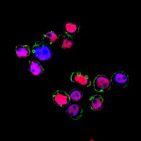
Immunocytochemistry Analysis: A representative lot detected MNDA nuclear immunoreactivity among HL-60 cells and NMDA-transfected, but not untransfected K562 cells. MNDA-negative HL-60 cells increased with time in cultures maintained without fresh medium for an extended period of time. MNDA translocated from nucleus to cytoplasm beginning at 3 hours and completed 4 hours following H2O2 addition to HL-60 cells (Briggs, R.C., et al. (2006). Cancer Res. 66(9):4645-4651).

Immunoprecipitation Analysis: A representative lot immunoprecipitated MNDA from HL-60 nuclear extract (Hudson, C.R., et al. (1988). Hybridoma. 7(6):541-553).

Western Blotting Analysis: A representative lot detected a time-dependent loss of MNDA in HL-60 cell lysate beginning at 4 hours following H2O2 treatment (Briggs, R.C., et al. (2006). Cancer Res. 66(9):4645-4651).

Western Blotting Analysis: Clone 3C1 hybridoma culture supernatant detected MNDA in HL-60 nuclear extract (Hudson, C.R., et al. (1988). Hybridoma. 7(6):541-553).
Biological Information
ImmunogenHuman peripheral blood granulocyte nuclei.
Clone3C1
ConcentrationPlease refer to lot specific datasheet.
HostRat
SpecificityClone 3C1 reacted with both denatured and non-denatured MNDA. Clone 3C1 reacted with nuclear extract from NMDA-expressing HL-60, but not non-MNDA-expressing BII, human myeloid leukemia cells (Hudson, C.R., et al. (1988). Hybridoma. 7(6):541-553).
IsotypeIgG1κ
Species Reactivity
  • Human
Species Reactivity NoteHuman.
Antibody TypeMonoclonal Antibody
Entrez Gene Number
Gene Symbol
  • MNDA
Purification MethodProtein G purified.
UniProt Number
Molecular Weight45.84 kDa calculated. ~55 kDa reported (Hudson, C.R., et al. (1988). Hybridoma. 7(6):541-553). Uncharacterized bands may be observed in some lysate(s).
Physicochemical Information
Dimensions
Materials Information
Toxicological Information
Safety Information according to GHS
Safety Information
Product Usage Statements
Quality AssuranceEvaluated by Immunocytochemistry in HL-60 cells.

Immunocytochemistry Analysis: A 1:1,000 dilution of this antibody detected MNDA in 4% paraformaldehyde-fixed, 0.3% Triton X-100-permeabilized HL-60 cells.
Usage Statement
  • Unless otherwise stated in our catalog or other company documentation accompanying the product(s), our products are intended for research use only and are not to be used for any other purpose, which includes but is not limited to, unauthorized commercial uses, in vitro diagnostic uses, ex vivo or in vivo therapeutic uses or any type of consumption or application to humans or animals.
Storage and Shipping Information
Storage ConditionsStable for 1 year at 2-8°C from date of receipt.
Packaging Information
Material Size100 μg
Transport Information
Supplemental Information
Specifications
Global Trade Item Number
Catalog Number GTIN
MABC1170 04054839090073