Crmp4 deletion promotes recovery from spinal cord injury by neuroprotection and limited scar formation.
Nagai, J; Kitamura, Y; Owada, K; Yamashita, N; Takei, K; Goshima, Y; Ohshima, T
Scientific reports
5
8269
2015
Show Abstract
Axonal outgrowth inhibitors and scar formation are two major obstacles to central nervous system (CNS) repair. No target molecule that regulates both axonal growth and scarring has been identified. Here we identified collapsin response mediator protein 4 (CRMP4), a common mediator of inhibitory signals after neural injury, as a crucial factor that contributes to both axonal growth inhibition and scarring after spinal cord injury (SCI). We found increases in the inhibitory and toxic forms of CRMP4 in injured spinal cord. Notably, CRMP4 expression was evident in inflammatory cells as well as in neurons after spinal cord transection. Crmp4-/- mice displayed neuroprotection against SCI and reductions in inflammatory response and scar formation. This permissive environment for axonal growth due to CRMP4 deletion restored locomotor activity at an unusually early phase of healing. These results suggest that deletion of CRMP4 is a unique therapeutic strategy that overcomes two obstacles to CNS repair after SCI. | Immunofluorescence | 25652774
 |
Immunopanning purification and long-term culture of human retinal ganglion cells.
Zhang, XM; Li Liu, DT; Chiang, SW; Choy, KW; Pang, CP; Lam, DS; Yam, GH
Molecular vision
16
2867-72
2010
Show Abstract
To establish a robust method to isolate primary retinal ganglion cells (RGCs) from human fetal retina for long-term culture while maintaining neuronal morphology and marker protein expression.A total of six human retinas were obtained from aborted fetuses at 10 to 12 weeks of gestation with informed consent from mothers. RGCs were isolated and purified by a modified two-step immunopanning procedure. The cells were maintained in a serum-free defined medium supplemented with brain-derived neurotrophic factor, ciliary neutrophic factor, and forskolin. The viable RGCs and the extent of neurite outgrowth were examined by calcein-acetoxymethylester assay. Expression of RGC markers was studied by immunocytochemistry.Primary RGCs from human fetal retinas were isolated and maintained in vitro for one month with substantial neurite elongation. In cell culture, almost 70% of the isolated cells attached, spread, and displayed numerous dendrites. They were immunoreactive to RGC-specific markers (Thy-1, TUJ-1, and Brn3a) and negative for glial fibrillary acidic protein and amacrine cells marker HPC-1.Human RGCs were successfully isolated and maintained in long-term culture. This can serve as an ideal model for biologic, toxicological, and genomic assays of human RGCs in vitro. Full Text Article | | 21203402
 |
The localization of populations of lymphocytes defined by monoclonal antibodies in rat lymphoid tissues.
Barclay, A N
Immunology, 42: 593-600 (1981)
1981
Show Abstract
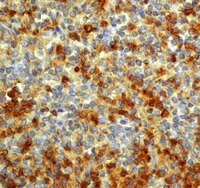
The localisation of subpopulations of T lymphocytes as defined by the monoclonal antibodies W3/25 and MRC OX 8 was determined by an indirect immunoperoxidase technique on cryostat sections of rat lymphoid tissues. Both subpopulations were present throughout T-dependent areas with only scattered T cells, largely W3/25 positive, in the B areas. The W3/25 antigen was also found on macrophages in the medulla of lymph nodes and in the peritoneal cavity and liver. | | 7016746
 |


