Functional role of neurotrophin-3 in synapse regeneration by spiral ganglion neurons on inner hair cells after excitotoxic trauma in vitro.
Wang, Q; Green, SH
The Journal of neuroscience : the official journal of the Society for Neuroscience
31
7938-49
2011
Show Abstract
Spiral ganglion neurons (SGNs) are postsynaptic to hair cells and project to the brainstem. The inner hair cell (IHC) to SGN synapse is susceptible to glutamate excitotoxicity and to acoustic trauma, with potentially adverse consequences to long-term SGN survival. We used a cochlear explant culture from P6 rat pups consisting of a portion of organ of Corti maintained intact with the corresponding portion of spiral ganglion to investigate excitotoxic damage to IHC-SGN synapses in vitro. The normal innervation pattern is preserved in vitro. Brief treatment with NMDA and kainate results in loss of IHC-SGN synapses and degeneration of the distal type 1 SGN peripheral axons, mimicking damage to SGN peripheral axons caused by excitotoxicity or noise in vivo. The number of IHC presynaptic ribbons is not significantly altered. Reinnervation of IHCs occurs and regenerating axons remain restricted to the IHC row. However, the number of postsynaptic densities (PSDs) does not fully recover and not all axons regrow to the IHCs. Addition of either neurotrophin-3 (NT-3) or BDNF increases axon growth and synaptogenesis. Selective blockade of endogenous NT-3 signaling with TrkC-IgG reduced regeneration of axons and PSDs, but TrkB-IgG, which blocks BDNF, has no such effect, indicating that endogenous NT-3 is necessary for SGN axon growth and synaptogenesis. Remarkably, TrkC-IgG reduced axon growth and synaptogenesis even in the presence of BDNF, indicating that endogenous NT-3 has a distinctive role, not mimicked by BDNF, in promoting SGN axon growth in the organ of Corti and synaptogenesis on IHCs. | 21613508
 |
Pathogenicity of HIV in lymphatic organs of patients with AIDS.
Pekovic, D D, et al.
J. Pathol., 152: 31-5 (1987)
1987
Show Abstract
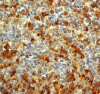
HIV antigens were searched for in the thymus, lymph nodes, bone marrow, and spleen of AIDS patients, by means of immunofluorescence technique. Human IgG against HIV and monoclonal antibodies against viral gag P24 protein yielded strong cytoplasmic fluorescence of cells in sections of the thymus, lymph nodes and spleen. Some cells containing HIV antigens were morphologically multinucleated giant cells. They reacted with monoclonal antibodies against helper/inducer T-cells (OKT4+), and were complexed with antibody or with complement as demonstrated by double-staining immunofluorescence technique. A large number of inflammatory cells infiltrated the thymus in areas containing cells expressing HIV antigens. These studies demonstrated an association of HIV virus with cytopathic and immunopathogenic reactions in lymphatic organs of AIDS patients, and are consistent with previous results, as well as indicative of a primary aetiologic role for the virus. | 3305846
 |


