Millipore Sigma Vibrant Logo
Attention: We have moved. EMD Millipore products are no longer available for purchase on emdmillipore.com.Learn More

07-1231 Anti-Notch1 Antibody, Intracellular

View Products on Sigmaaldrich.com
07-1231
200 µL  
Purchase on Sigma-Aldrich

Special Offers

Overview

Replacement Information

Key Specifications Table

Species ReactivityKey ApplicationsHostFormatAntibody Type
HELISA, WB, IH(P)RbSerumPolyclonal Antibody
Description
Catalogue Number07-1231
Replaces04-1046
DescriptionAnti-Notch1 Antibody, Intracellular
Alternate Names
  • Neurogenic locus notch homolog protein 1 precursor
  • Notch (Drosophila) homolog 1 (translocation-associated)
  • Notch 1
  • Notch homolog 1, translocation-associated (Drosophila)
  • Translocation-associated notch protein TAN-1
  • Neurogenic locus notch homolog protein 1
Background InformationNotch is a family of single-pass transmembrane receptor proteins that are synthesized in the endoplasmic reticulum as an inactive form and are proteolytically cleaved on an extracellular site by a furin-like convertase (S1 cleavage) in the trans-golgi network after the recognition of the RQRR sequence before it reaches the plasma membrane as heterodimers to yield an active, ligand-accessible form. The Notch family is comprised of 4 members (1-4) whose ligands include the Delta and Jagged family of ligands. These ligands cause proteolysis of Notch to liberate the intracellular domain. Cleavage results in a C-terminal fragment N(TN) and a N-terminal fragment N(EC). Following ligand binding, it is cleaved (S2 cleavage) by TNF-alpha converting enzyme (TACE) to yield a membrane-associated intermediate fragment called Notch extracellular truncation (NEXT). This fragment is then cleaved by presenilin-dependent gamma-secretase between gly1743 and val1744 (S3 cleavage) to release the intracellular domain (NICD) from the membrane. That domain translocates to the nucleus and initiates transcription events by binding the DNA binding protein CSL. The notch family members are involved cell differentiation and development.
References
Product Information
FormatSerum
HS Code3002 15 90
Control
  • HeLa Cell Lysate
PresentationRabbit polyclonal IgG serum in buffer containing in 0.02 M Potassium phosphate, 0.15 M NaCl with 0.01% sodium azide.
Quality LevelMQ100
Applications
ApplicationUse Anti-Notch1 Antibody, Intracellular (Rabbit Polyclonal Antibody) validated in ELISA, WB, IHC(P) to detect Notch1 also known as Neurogenic locus notch homolog protein 1 precursor.
Key Applications
  • ELISA
  • Western Blotting
  • Immunohistochemistry (Paraffin)
Application NotesELISA: 1:20,000-1:100,000 dilution from a previous lot was used in a standard sandwich ELISA assay against the peptide immunogen.
Optimal working dilutions must be determined by the end user.

Immunohistochemistry(paraffin):
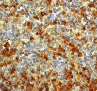
Representative testing from a previous lot.
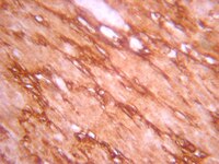
Optimal Staining IGF-1 With Epitope Retrieval: Lipoma.
Biological Information
ImmunogenSynthetic peptide from human Notch 1.
EpitopeIntracellular
HostRabbit
SpecificityNotch 1, intracellular. No reactivity against human Notch 2.
IsotypeIgG
Species Reactivity
  • Human
Species Reactivity NoteHuman. No reactivity observed to mouse Notch. Reactivity with other species has not been determined.
Antibody TypePolyclonal Antibody
Entrez Gene Number
Entrez Gene SummaryThis gene encodes a member of the Notch family. Members of this Type 1 transmembrane protein family share structural characteristics including an extracellular domain consisting of multiple epidermal growth factor-like (EGF) repeats, and an intracellular domain consisting of multiple, different domain types. Notch family members play a role in a variety of developmental processes by controlling cell fate decisions. The Notch signaling network is an evolutionarily conserved intercellular signaling pathway which regulates interactions between physically adjacent cells. In Drosophilia, notch interaction with its cell-bound ligands (delta, serrate) establishes an intercellular signaling pathway that plays a key role in development. Homologues of the notch-ligands have also been identified in human, but precise interactions between these ligands and the human notch homologues remain to be determined. This protein is cleaved in the trans-Golgi network, and presented on the cell surface as a heterodimer. This protein functions as a receptor for membrane bound ligands, and may play multiple roles during development.
Gene Symbol
  • NOTCH1
  • notch1
  • hN1
  • TAN1
UniProt Number
UniProt SummaryFUNCTION: SwissProt: P46531 # Functions as a receptor for membrane-bound ligands Jagged1, Jagged2 and Delta1 to regulate cell-fate determination. Upon ligand activation through the released notch intracellular domain (NICD) it forms a transcriptional activator complex with RBP-J kappa and activates genes of the enhancer of split locus. Affects the implementation of differentiation, proliferation and apoptotic programs. May be important for normal lymphocyte function. In altered form, may contribute to transformation or progression in some T-cell neoplasms. Involved in the maturation of both CD4+ and CD8+ cells in the thymus. May be important for follicular differentiation and possibly cell fate selection within the follicle. During cerebellar development, may function as a receptor for neuronal DNER and may be involved in the differentiation of Bergmann glia (By similarity).
SIZE: 2556 amino acids; 272500 Da
SUBUNIT: Heterodimer of a C-terminal fragment N(TM) and an N- terminal fragment N(EC) which are probably linked by disulfide bonds. Interacts with DNER, DTX1, DTX2 and RBPSUH. Also interacts with MAML1, MAML2 and MAML3 which act as transcriptional coactivators for NOTCH1.
SUBCELLULAR LOCATION: Cell membrane; Single-pass type I membrane protein (By similarity). & Notch 1 intracellular domain: Nucleus (By similarity). Note=Following proteolytical processing NICD is translocated to the nucleus (By similarity).
TISSUE SPECIFICITY: In fetal tissues most abundant in spleen, brain stem and lung. Also present in most adult tissues where it is found mainly in lymphoid tissues.
PTM: Synthesized in the endoplasmic reticulum as an inactive form which is proteolytically cleaved by a furin-like convertase in the trans-Golgi network before it reaches the plasma membrane to yield an active, ligand-accessible form. Cleavage results in a C- terminal fragment N(TM) and a N-terminal fragment N(EC). Following ligand binding, it is cleaved by TNF-alpha converting enzyme (TACE) to yield a membrane-associated intermediate fragment called notch extracellular truncation (NEXT). This fragment is then cleaved by presenilin dependent gamma-secretase to release a notch-derived peptide containing the intracellular domain (NICD) from the membrane (By similarity). & Phosphorylated (By similarity).
DISEASE: SwissProt: P46531 # NOTCH1 truncation is associated with T-cell acute lymphoblastic leukemia. & Defects in NOTCH1 are a cause of aortic valve disease [MIM:109730]. The disorder consists of an early developmental defect in the aortic valve and a later de-repression of calcium deposition that causes progressive aortic valve disease. Calcification of the aortic valve is the third leading cause of heart disease in adults. The incidence increases with age, and it is often associated with a bicuspid aortic valve present in 1-2% of the population.
SIMILARITY: SwissProt: P46531 ## Belongs to the NOTCH family. & Contains 5 ANK repeats. & Contains 36 EGF-like domains. & Contains 3 LNR (Lin/Notch) repeats.
Molecular Weight~80 kDa
Physicochemical Information
Dimensions
Materials Information
Toxicological Information
Safety Information according to GHS
Safety Information
Product Usage Statements
Quality AssuranceRoutinely evaluated by Western Blot on HeLa lysates.

Western Blot Analysis: 1:500 dilution of this lot detected NOTCH1, intracellular (Cleaved form) on 10 μg of HeLa lysates.
Usage Statement
  • Unless otherwise stated in our catalog or other company documentation accompanying the product(s), our products are intended for research use only and are not to be used for any other purpose, which includes but is not limited to, unauthorized commercial uses, in vitro diagnostic uses, ex vivo or in vivo therapeutic uses or any type of consumption or application to humans or animals.
Storage and Shipping Information
Storage ConditionsStable for 6 months at -20°C in undiluted aliquots from date of receipt. Avoid repeated freeze/thaw cycles.
Handling Recommendations: Upon first thaw, and prior to removing the cap, centrifuge the vial and gently mix the solution. Aliquot into microcentrifuge tubes and store at -20°C. Avoid repeated freeze/thaw cycles, which may damage IgG and affect product performance.
Packaging Information
Material Size200 µL
Transport Information
Supplemental Information
Specifications
Global Trade Item Number
Catalog Number GTIN
07-1231 04053252516528

Documentation

Anti-Notch1 Antibody, Intracellular SDS

Title

Safety Data Sheet (SDS) 

Anti-Notch1 Antibody, Intracellular Certificates of Analysis

TitleLot Number
Anti-Notch 1, Intracellular 2842910
Anti-Notch 1, Intracellular - 2410224 2410224
Anti-Notch 1, Intracellular - 2018493 2018493
Anti-Notch 1, Intracellular - 3190920 3190920
Anti-Notch 1, Intracellular - 3485905 3485905
Anti-Notch 1, Intracellular - 3506695 3506695
Anti-Notch 1, Intracellular - 3845722 3845722
Anti-Notch 1, Intracellular - 3932194 3932194
Anti-Notch 1, Intracellular - 4055465 4055465
Anti-Notch 1, Intracellular - 4073640 4073640

Related Products & Applications

Related Products

Catalog Number Description
07-1232 Anti-Notch 1 Antibody, NT
07-218 Anti-Notch1 Antibody, extracellular domain
MAB5352 Anti-Notch 1 Antibody, clone mN1A
MAB5414 Anti-Notch 1 Antibody, extracellular, clone 8G10

Product Families

Categories

Life Science Research > Antibodies and Assays > Primary Antibodies