Millipore Sigma Vibrant Logo
Attention: We have moved. EMD Millipore products are no longer available for purchase on emdmillipore.com.Learn More

MABF261 Anti-PD-1 Antibody, clone 16A2.1

View Products on Sigmaaldrich.com
MABF261
100 μg  
Purchase on Sigma-Aldrich

Special Offers

Overview

Replacement Information

Key Specifications Table

Species ReactivityKey ApplicationsHostFormatAntibody Type
HWB, FCMAffinity PurifiedMonoclonal Antibody
Description
Catalogue NumberMABF261
DescriptionAnti-PD-1 Antibody, clone 16A2.1
Alternate Names
  • PD-1
  • Programmed cell death protein 1
  • Protein PD-1
  • hPD-1
  • CD279
Background InformationProgrammed cell death protein 1 (PD-1) (UniProt Q15116), also known as CD279, hPD-1, hPD-I, hSLE1, Protein PD-1, Systemic lupus erythematosus susceptibility 2) is a type I single transmembrane protein encoded by the PDCD1 gene in human. Maintenance of peripheral tolerance by preventing the development of autoimmunity due to T lymphocytes specific for self-antigens is essential for homeostasis of the immune system. PD-1 receptor CD279 and its ligands PD-L1 (B7-H1, CD274) and PD-L2 (B7-DC; CD273) play a vital role in the induction and maintenance of peripheral tolerance. Upregulated PD-1 expression is seen on conventional CD4+ CD25- T cells (Tconv) when exposed to mesenchymal stem cells, resulting in increased Tconv apoptosis due to PD-1 stimulation by the MSC PD-L2.
References
Product Information
FormatAffinity Purified
PresentationPurified mouse monoclonal IgG2ak in buffer containing 0.1 M Tris-Glycine (pH 7.4), 150 mM NaCl with 0.05% sodium azide.
Quality LevelMQ100
Applications
ApplicationAnti-PD-1 Antibody, clone 16A2.1 is an antibody against PD-1 for use in Western Blotting, Flow Cytometry.
Key Applications
  • Western Blotting
  • Flow Cytometry
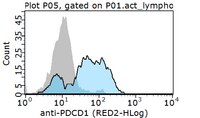
Application NotesFlow Cytometry Analysis: 2.0 µg of this antibody from a representative lot detected PD-1 in 1X10E6 Staph enterotoxin B treated human peripheral blood mononuclear cells.
Biological Information
ImmunogenKLH-conjugated linear peptide corresponding to the extracellular domain of human PD-1.
EpitopeExtracellular domain
Clone16A2.1
ConcentrationPlease refer to lot specific datasheet.
HostMouse
IsotypeIgG2aκ
Species Reactivity
  • Human
Antibody TypeMonoclonal Antibody
Entrez Gene Number
Gene Symbol
  • PDCD1
  • PD1
Purification MethodProtein G Purified
UniProt Number
Molecular Weight~32 kDa observed
Physicochemical Information
Dimensions
Materials Information
Toxicological Information
Safety Information according to GHS
Safety Information
Product Usage Statements
Quality AssuranceEvaluated by Western Blotting in TPA/LPS treated THP-1 cell lysate.

Western Blotting Analysis: 1.0 µg/mL of this antibody detected PD-1 in 10 µg of TPA/LPS treated THP-1 cell lysate.
Usage Statement
  • Unless otherwise stated in our catalog or other company documentation accompanying the product(s), our products are intended for research use only and are not to be used for any other purpose, which includes but is not limited to, unauthorized commercial uses, in vitro diagnostic uses, ex vivo or in vivo therapeutic uses or any type of consumption or application to humans or animals.
Storage and Shipping Information
Storage ConditionsStable for 1 year at 2-8°C from date of receipt.
Packaging Information
Material Size100 μg
Transport Information
Supplemental Information
Specifications
Global Trade Item Number
Catalog Number GTIN
MABF261 04055977168280

Documentation

Anti-PD-1 Antibody, clone 16A2.1 Certificates of Analysis

TitleLot Number
Anti-PD-1, clone 16A2.1 - 3478265 3478265
Anti-PD-1, clone 16A2.1 - 4082281 4082281
Anti-PD-1, clone 16A2.1 -Q2569101 Q2569101
Anti-PD-1, clone 16A2.1 Monoclonal Antibody 2929105
Anti-PD-1, clone 16A2.1 Monoclonal Antibody 3009966

Technical Info

Title
Characterization of Estrogen Receptor α Phosphorylation Sites in Breast Cancer Tissue Using the SNAP i.d® 2.0 System
White Paper: Further considerations of antibody validation and usage.