Epitope mapping of anti-PGRMC1 antibodies reveals the non-conventional membrane topology of PGRMC1 on the cell surface
Ji Yea Kim 1 2 , So Young Kim 1 , Hong Seo Choi 1 , Sungkwan An 2 , Chun Jeih Ryu
Sci Rep
9(1)
653
2019
Show Abstract
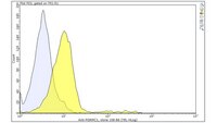
Progesterone receptor membrane component1 (PGRMC1) is a heme-binding protein involved in cancers and Alzheimer's disease. PGRMC1 consists of a short N-terminal extracellular or luminal domain, a single membrane-spanning domain, and a long cytoplasmic domain. Previously, we generated two monoclonal antibodies (MAbs) 108-B6 and 4A68 that recognize cell surface-expressed PGRMC1 (csPGRMC1) on human pluripotent stem cells and some cancer cells. In this study, flow cytometric analysis found that an anti-PGRMC1 antibody recognizing the N-terminus of PGRMC1 could not bind to csPGRMC1 on cancer cells, and 108-B6 and 4A68 binding to csPGRMC1 was inhibited by trypsin treatment, suggesting that the epitopes of 108-B6 and 4A68 are trypsin-sensitive. To examine the epitope specificity of 108-B6 and 4A68, glutathione-S-transferase (GST)-fused PGRMC1 mutants were screened to identify the epitopes targeted by the antibodies. The result showed that 108-B6 and 4A68 recognized C-terminal residues 183-195 and 171-182, respectively, of PGRMC1, where trypsin-sensitive sites are located. A polyclonal anti-PGRMC1 antibody raised against the C-terminus of PGRMC1 could also recognized csPGRMC1 in a trypsin-sensitive manner, suggesting that the C-terminus of csPGRMC1 is exposed on the cell surface. This finding reveals that csPGRMC1 has a non-conventional plasma membrane topology, which is different from that of intracellular PGRMC1. | 30679694
 |
Progesterone Receptor Membrane Component 1 suppresses the p53 and Wnt/β-catenin pathways to promote human pluripotent stem cell self-renewal
Ji Yea Kim 1 , So Young Kim 1 , Hong Seo Choi 1 , Min Kyu Kim 1 , Hyun Min Lee 1 , Young-Joo Jang 2 , Chun Jeih Ryu
Sci Rep
8(1)
3048
2018
Show Abstract
Progesterone receptor membrane component 1 (PGRMC1) is a multifunctional heme-binding protein involved in various diseases, including cancers and Alzheimer's disease. Previously, we generated two monoclonal antibodies (MAbs) 108-B6 and 4A68 against surface molecules on human pluripotent stem cells (hPSCs). Here we show that PGRMC1 is the target antigen of both MAbs, and is predominantly expressed on hPSCs and some cancer cells. PGRMC1 is rapidly downregulated during early differentiation of hPSCs. Although PGRMC1 knockdown leads to a spread-out morphology and impaired self-renewal in hPSCs, PGRMC1 knockdown hPSCs do not show apoptosis and autophagy. Instead, PGRMC1 knockdown leads to differentiation of hPSCs into multiple lineage cells without affecting the expression of pluripotency markers. PGRMC1 knockdown increases cyclin D1 expression and decreases Plk1 expression in hPSCs. PGRMC1 knockdown also induces p53 expression and stability, suggesting that PGRMC1 maintains hPSC self-renewal through suppression of p53-dependent pathway. Analysis of signaling molecules further reveals that PGRMC1 knockdown promotes inhibitory phosphorylation of GSK-3β and increased expression of Wnt3a and β-catenin, which leads to activation of Wnt/β-catenin signaling. The results suggest that PGRMC1 suppresses the p53 and Wnt/β-catenin pathways to promote self-renewal and inhibit early differentiation in hPSCs. | 29445107
 |
Development of a decoy immunization strategy to identify cell-surface molecules expressed on undifferentiated human embryonic stem cells
Hong Seo Choi 1 , Hana Kim, Ayoung Won, Jum-Ji Kim, Chae-Yeon Son, Kyoung-Soo Kim, Jeong Heon Ko, Mi-Young Lee, Cheorl-Ho Kim, Chun Jeih Ryu
Cell Tissue Res
333(2)
197-206
2008
Show Abstract
Little is known about the cell-surface molecules that are related to the undifferentiated and pluripotent state of human embryonic stem cells (hESCs). Here, we generated a panel of murine monoclonal antibodies (MAb) against undifferentiated hESCs by a modification of a previously described decoy immunization strategy. H9 hESCs were differentiated in the presence of retinoic acid and used as a decoy immunogen. Twelve Balb/c mice were immunized in the right hind footpads with differentiated H9 cells and in the left hind footpads with undifferentiated H9 cells. After immunization, the left popliteal lymph node cells were collected and were fused with mouse myeloma cells. The fusion resulted in 79 hybridomas secreting MAbs that bound to the undifferentiated H9 cells as shown by flow cytometric analysis. Of these, 70 MAbs bound to the undifferentiated H9 cells, but only weakly or not at all to the differentiated H9 cells. We characterized 37 MAbs (32 IgGs, 5 IgMs) recognizing surface molecules that were down-regulated during embryoid body cell formation. One of the MAbs, L125-C2, was confirmed to immunoprecipitate CD9, previously known as a surface molecule on the undifferentiated hESCs. To investigate the relationship between the MAbs and hESC-specific antibodies, two representative MAbs, viz., L125-C2 and 291-D4, were selected and studied by multi-color flow cytometric analysis. This showed that more than 60% of L125-C2- and 291-D4-positive cells were also positive for the expression of hESC-specific surface molecules such as SSEA3, SSEA4, TRA-1-60, and TRA-1-81, indicating the close relationship between the two MAbs and the hESC-specific surface molecules. Our results suggest that the decoy immunization strategy is an efficient method for isolating a panel of MAbs against undifferentiated hESCs, and that the generated MAbs should be useful for studying the surface molecules on hESCs in the pluripotent and undifferentiated state. | 18560898
 |


