Millipore Sigma Vibrant Logo
Attention: We have moved. EMD Millipore products are no longer available for purchase on emdmillipore.com.Learn More

MABS2041-100UG Anti-pan-ARF Antibody, clone 1D9

View Products on Sigmaaldrich.com
MABS2041-100UG
100 μg  
Purchase on Sigma-Aldrich

Overview

Replacement Information

Key Specifications Table

Species ReactivityKey ApplicationsHostFormatAntibody Type
HELISA, EM, IP, WBMPurifiedMonoclonal Antibody
Description
Catalogue NumberMABS2041-100UG
DescriptionAnti-pan-ARF Antibody, clone 1D9
Alternate Names
  • ADP-ribosylation factor 1
Background InformationADP-ribosylation factor 1 (UniProt: P84077; also known as Arf1) is encoded by the ARF1 gene (Gene ID: 375) in human. Arf1 is a member of the family of low molecular weight GTP-binding proteins. It is abundant in neural tissues where it may comprise up to 1% of total cellular protein. The ARF proteins are categorized as class I (ARF1, ARF2 and ARF3), class II (ARF4 and ARF5) and class III (ARF6), and members of each class share a common gene organization. Arf1, 3, 4, and 5 are predominantly cytosolic, but could be recruited to a variety of intracellular membranes, but not plasma membranes, upon incubation in the presence of GTP S. Arf6 is found at the plasma membrane and in endosomes. Arf1 is localized to the Golgi apparatus and has a central role in intra-Golgi transport. It has two nucleotide binding regions (aa 24-32 and 126-129). Arf1 functions as an allosteric activator of the cholera toxin catalytic subunit, an ADP-ribosyltransferase. It is also involved in protein trafficking among different compartments and is reported to modulate vesicle budding and uncoating within the Golgi complex. In its GTP-bound form, its triggers the association with coat proteins with the Golgi membrane. The hydrolysis of Arf1-bound GTP, which is mediated by ARFGAPs proteins, is required for dissociation of coat proteins from Golgi membranes and vesicles. This antibody (clone 1D9) detects all Arf proteins, but to different degrees. (Ref.: Cavenagh, MM., et al. (1996). J. Biol. Chem. 271(36):21767-74).
References
Product Information
FormatPurified
PresentationPurified mouse monoclonal antibody IgG1 in buffer containing 0.1 M Tris-Glycine (pH 7.4), 150 mM NaCl with 0.05% sodium azide.
Quality LevelMQ100
Applications
ApplicationAnti-pan-ARF, clone 1D9, Cat. No. MABS2041, is a mouse monoclonal antibody that detects all ADP-ribosylation factors and has been tested for use in ELISA, Electron Microscopy, Immunoprecipitation, and Western Blotting,
Key Applications
  • ELISA
  • Electron Microscopy
  • Immunoprecipitation
  • Western Blotting
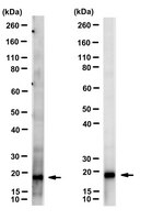
Application NotesWestern Blotting Analysis: 1 ug/mL from a representative lot detected ARF proteins in human lung tissue lysate.

Immunoprecipitation Analysis: A representative lot immunoprecipitated ARF proteins (Cavenagh, M.M., et. al. (1996). J Biol Chem. 271(36):21767-74).

ELISA Analysis: A representative lot detected pan-ARF in ELISA applications (Cavenagh, M.M., et. al. (1996). J Biol Chem. 271(36):21767-74).

Western Blotting Analysis: A representative lot detected ARF proteins in Western Blotting applications (Cavenagh, M.M., et. al. (1996). J Biol Chem. 271(36):21767-74; Zuezem, S., et. al. (1992). Proc Natl Acad Sci USA. 89(14):6619-23).

Electron Microscopy Analysis: A representative lot detected ARF proteins in Electron Microscopy applications (Zuezem, S., et. al. (1992). Proc Natl Acad Sci USA. 89(14):6619-23).

Dot Blot Analysis: A representative lot detected ARF proteins in Dot Blot applications (Cavenagh, M.M., et. al. (1996). J Biol Chem. 271(36):21767-74).
Biological Information
ImmunogenPurified full length human recombinant ADP-ribosylation factor 1.
Clone1D9
ConcentrationPlease refer to lot specific datasheet.
HostMouse
SpecificityClone 1D9 detects all isoforms of DP-ribosylation factors in human cells.
IsotypeIgG1κ
Species Reactivity
  • Human
Species Reactivity NoteHuman.
Antibody TypeMonoclonal Antibody
Entrez Gene Number
Gene Symbol
  • ARF1
Purification MethodProtein G purified
UniProt Number
Molecular Weight~20 kDa observed. Uncharacterized bands may be observed in some lysate(s).
Physicochemical Information
Dimensions
Materials Information
Toxicological Information
Safety Information according to GHS
Safety Information
Product Usage Statements
Quality AssuranceEvaluated by Western Blotting in HeLa cell lysate.

Western Blotting Analysis: 1 ug/mL of this antibody detected pan-ARF in HeLa cell lysate.
Usage Statement
  • Unless otherwise stated in our catalog or other company documentation accompanying the product(s), our products are intended for research use only and are not to be used for any other purpose, which includes but is not limited to, unauthorized commercial uses, in vitro diagnostic uses, ex vivo or in vivo therapeutic uses or any type of consumption or application to humans or animals.
Storage and Shipping Information
Storage ConditionsStable for 1 year at 2-8°C from date of receipt.
Packaging Information
Material Size100 μg
Transport Information
Supplemental Information
Specifications
Global Trade Item Number
Catalog Number GTIN
MABS2041-100UG 04054839448294