Dual targeting of microtubule and topoisomerase II by α-carboline derivative YCH337 for tumor proliferation and growth inhibition.
Yi, JM; Zhang, XF; Huan, XJ; Song, SS; Wang, W; Tian, QT; Sun, YM; Chen, Y; Ding, J; Wang, YQ; Yang, CH; Miao, ZH
Oncotarget
6
8960-73
2015
Show Abstract
Both microtubule and topoisomerase II (Top2) are important anticancer targets and their respective inhibitors are widely used in combination for cancer therapy. However, some combinations could be mutually antagonistic and drug resistance further limits their therapeutic efficacy. Here we report YCH337, a novel α-carboline derivative that targets both microtubule and Top2, eliciting tumor proliferation and growth inhibition and overcoming drug resistance. YCH337 inhibited microtubule polymerization by binding to the colchicine site and subsequently led to mitotic arrest. It also suppressed Top2 and caused DNA double-strand breaks. It disrupted microtubule more potently than Top2. YCH337 induced reversible mitotic arrest at low concentrations but persistent DNA damage. YCH337 caused intrinsic and extrinsic apoptosis and decreased MCL-1, cIAP1 and XIAP proteins. In this aspect, YCH337 behaved differently from the combination of vincristine and etoposide. YCH337 inhibited proliferation of tumor cells with an averaged IC50 of 0.3 μM. It significantly suppressed the growth of HT-29 xenografts in nude mice too. Importantly, YCH337 nearly equally killed different-mechanism-mediated resistant tumor cells and corresponding parent cells. Together with the novelty of its chemical structure, YCH337 could serve as a promising lead for drug development and a prototype for a dual microtubule/Top2 targeting strategy for cancer therapy. | | | 25840421
 |
FBXW7 and USP7 regulate CCDC6 turnover during the cell cycle and affect cancer drugs susceptibility in NSCLC.
Morra, F; Luise, C; Merolla, F; Poser, I; Visconti, R; Ilardi, G; Paladino, S; Inuzuka, H; Guggino, G; Monaco, R; Colecchia, D; Monaco, G; Cerrato, A; Chiariello, M; Denning, K; Claudio, PP; Staibano, S; Celetti, A
Oncotarget
6
12697-709
2015
Show Abstract
CCDC6 gene product is a pro-apoptotic protein substrate of ATM, whose loss or inactivation enhances tumour progression. In primary tumours, the impaired function of CCDC6 protein has been ascribed to CCDC6 rearrangements and to somatic mutations in several neoplasia. Recently, low levels of CCDC6 protein, in NSCLC, have been correlated with tumor prognosis. However, the mechanisms responsible for the variable levels of CCDC6 in primary tumors have not been described yet.We show that CCDC6 turnover is regulated in a cell cycle dependent manner. CCDC6 undergoes a cyclic variation in the phosphorylated status and in protein levels that peak at G2 and decrease in mitosis. The reduced stability of CCDC6 in the M phase is dependent on mitotic kinases and on degron motifs that are present in CCDC6 and direct the recruitment of CCDC6 to the FBXW7 E3 Ubl. The de-ubiquitinase enzyme USP7 appears responsible of the fine tuning of the CCDC6 stability, affecting cells behaviour and drug response.Thus, we propose that the amount of CCDC6 protein in primary tumors, as reported in lung, may depend on the impairment of the CCDC6 turnover due to altered protein-protein interaction and post-translational modifications and may be critical in optimizing personalized therapy. | | | 25885523
 |
Multiple assembly mechanisms anchor the KMN spindle checkpoint platform at human mitotic kinetochores.
Kim, S; Yu, H
The Journal of cell biology
208
181-96
2015
Show Abstract
During mitosis, the spindle checkpoint senses kinetochores not properly attached to spindle microtubules and prevents precocious sister-chromatid separation and aneuploidy. The constitutive centromere-associated network (CCAN) at inner kinetochores anchors the KMN network consisting of Knl1, the Mis12 complex (Mis12C), and the Ndc80 complex (Ndc80C) at outer kinetochores. KMN is a critical kinetochore receptor for both microtubules and checkpoint proteins. Here, we show that nearly complete inactivation of KMN in human cells through multiple strategies produced strong checkpoint defects even when all kinetochores lacked microtubule attachment. These KMN-inactivating strategies reveal multiple KMN assembly mechanisms at human mitotic kinetochores. In one mechanism, the centromeric kinase Aurora B phosphorylates Mis12C and strengthens its binding to the CCAN subunit CENP-C. In another, CENP-T contributes to KMN attachment in a CENP-H-I-K-dependent manner. Our study provides insights into the mechanisms of mitosis-specific assembly of the checkpoint platform KMN at human kinetochores. | | | 25601404
 |
Protein kinase C β inhibition by enzastaurin leads to mitotic missegregation and preferential cytotoxicity toward colorectal cancer cells with chromosomal instability (CIN).
Ouaret, D; Larsen, AK
Cell cycle (Georgetown, Tex.)
13
2697-706
2014
Show Abstract
Enzastaurin is a selective inhibitor of protein kinase C β and a potent inhibitor of tumor angiogenesis. In addition, enzastaurin shows direct cytotoxic activity toward a subset of tumor cells including colorectal cancer cells (CRC). In spite of promising results in animal models, the clinical activity of enzastaurin in CRC patients has been disappointing although a subset of patients seems to derive benefit. In the present study we investigated the biological and cytotoxic activities of enzastaurin toward a panel of well-characterized CRC cell lines in order to clarify the mechanistic basis for the cytotoxic activity. Our results show that enzastaurin is significantly more cytotoxic toward CRC cells with chromosome instability (CIN) compared to cells with microsatellite instability (MSI). Since CIN is usually attributed to mitotic dysfunction, the influence of enzastaurin on cell cycle progression and mitotic transit was characterized for representative CIN and MSI cell lines. Enzastaurin exposure was accompanied by prolonged metaphase arrest in CIN cells followed by the appearance of tetraploid and micronuclei-containing cells as well as by increased apoptosis, whereas no detectable mitotic dysfunctions were observed in MSI cells exposed to isotoxic doses of enzastaurin. Our study identifies enzastaurin as a new, context dependent member of a heterogeneous group of anticancer compounds that induce "mitotic catastrophe," that is mitotic dysfunction accompanied by cell death. These data provide novel insight into the mechanism of action of enzastaurin and may allow the identification of biomarkers useful to identify CRC patients particularly likely, or not, to benefit from treatment with enzastaurin. | | | 25486357
 |
Identification of a mitotic death signature in cancer cell lines.
Sakurikar, N; Eichhorn, JM; Alford, SE; Chambers, TC
Cancer letters
343
232-8
2014
Show Abstract
This study examined the molecular mechanism of action of anti-mitotic drugs. The hypothesis was tested that death in mitosis occurs through sustained mitotic arrest with robust Cdk1 signaling causing complete phosphorylation of Mcl-1 and Bcl-xL, and conversely, that mitotic slippage is associated with incomplete phosphorylation of Mcl-1/Bcl-xL. The results, obtained from studying six different cancer cell lines, strongly support the hypothesis and identify for the first time a unique molecular signature for mitotic death. The findings represent an important advance in understanding anti-mitotic drug action and provide insight into cancer cell susceptibility to such drugs which has important clinical implications. | | | 24099917
 |
Homeostatic control of polo-like kinase-1 engenders non-genetic heterogeneity in G2 checkpoint fidelity and timing.
Liang, H; Esposito, A; De, S; Ber, S; Collin, P; Surana, U; Venkitaraman, AR
Nature communications
5
4048
2014
Show Abstract
The G2 checkpoint monitors DNA damage, preventing mitotic entry until the damage can be resolved. The mechanisms controlling checkpoint recovery are unclear. Here, we identify non-genetic heterogeneity in the fidelity and timing of damage-induced G2 checkpoint enforcement in individual cells from the same population. Single-cell fluorescence imaging reveals that individual damaged cells experience varying durations of G2 arrest, and recover with varying levels of remaining checkpoint signal or DNA damage. A gating mechanism dependent on polo-like kinase-1 (PLK1) activity underlies this heterogeneity. PLK1 activity continually accumulates from initial levels in G2-arrested cells, at a rate inversely correlated to checkpoint activation, until it reaches a threshold allowing mitotic entry regardless of remaining checkpoint signal or DNA damage. Thus, homeostatic control of PLK1 by the dynamic opposition between checkpoint signalling and pro-mitotic activities heterogeneously enforces the G2 checkpoint in each individual cell, with implications for cancer pathogenesis and therapy. | Immunofluorescence | | 24893992
 |
Orally active microtubule-targeting agent, MPT0B271, for the treatment of human non-small cell lung cancer, alone and in combination with erlotinib.
Tsai, AC; Wang, CY; Liou, JP; Pai, HC; Hsiao, CJ; Chang, JY; Wang, JC; Teng, CM; Pan, SL
Cell death & disease
5
e1162
2014
Show Abstract
Microtubule-binding agents, such as taxanes and vinca alkaloids, are used in the treatment of cancer. The limitations of these treatments, such as resistance to therapy and the need for intravenous administration, have encouraged the development of new agents. MPT0B271 (N-[1-(4-Methoxy-benzenesulfonyl)-2,3-dihydro-1H-indol-7-yl]-1-oxy-isonicotinamide), an orally active microtubule-targeting agent, is a completely synthetic compound that possesses potent anticancer effects in vitro and in vivo. Tubulin polymerization assay and immunofluorescence experiment showed that MPT0B271 caused depolymerization of tubulin at both molecular and cellular levels. MPT0B271 reduced cell growth and viability at nanomolar concentrations in numerous cancer cell lines, including a multidrug-resistant cancer cell line NCI/ADR-RES. Further studies indicated that MPT0B271 is not a substrate of P-glycoprotein (P-gp), as determined by flow cytometric analysis of rhodamine-123 (Rh-123) dye efflux and the calcein acetoxymethyl ester (calcein AM) assay. MPT0B271 also caused G2/M cell-cycle arrest, accompanied by the up-regulation of cyclin B1, p-Thr161 Cdc2/p34, serine/threonine kinases polo-like kinase 1, aurora kinase A and B and the downregulation of Cdc25C and p-Tyr15 Cdc2/p34 protein levels. The appearance of MPM2 and the nuclear translocation of cyclin B1 denoted M phase arrest in MPT0B271-treated cells. Moreover, MPT0B271 induced cell apoptosis in a concentration-dependent manner; it also reduced the expression of Bcl-2, Bcl-xL, and Mcl-1 and increased the cleavage of caspase-3 and -7 and poly (ADP-ribose) polymerase (PARP). Finally, this study demonstrated that MPT0B271 in combination with erlotinib significantly inhibits the growth of the human non-small cell lung cancer A549 cells as compared with erlotinib treatment alone, both in vitro and in vivo. These findings identify MPT0B271 as a promising new tubulin-binding compound for the treatment of various cancers. | | | 24722287
 |
Comparative analysis of mitosis-specific antibodies for bulk purification of mitotic populations by fluorescence-activated cell sorting.
Campbell, Amy E, et al.
BioTechniques, 56: 90-4 (2014)
2014
Show Abstract
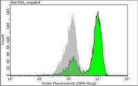
Mitosis entails complex chromatin changes that have garnered increasing interest from biologists who study genome structure and regulation-fields that are being advanced by high-throughput sequencing (Seq) technologies. The application of these technologies to study the mitotic genome requires large numbers of highly pure mitotic cells, with minimal contamination from interphase cells, to ensure accurate measurement of phenomena specific to mitosis. Here, we optimized a fluorescence-activated cell sorting (FACS)-based method for isolating formaldehyde-fixed mitotic cells-at virtually 100% mitotic purity and in quantities sufficient for high-throughput genomic studies. We compared several commercially available antibodies that react with mitosis-specific epitopes over a range of concentrations and cell numbers, finding antibody MPM2 to be the most robust and cost-effective. | Flow Cytometry | | 24502799
 |
Pin1-dependent signalling negatively affects GABAergic transmission by modulating neuroligin2/gephyrin interaction.
Antonelli, R; Pizzarelli, R; Pedroni, A; Fritschy, JM; Del Sal, G; Cherubini, E; Zacchi, P
Nature communications
5
5066
2014
Show Abstract
The cell adhesion molecule Neuroligin2 (NL2) is localized selectively at GABAergic synapses, where it interacts with the scaffolding protein gephyrin in the post-synaptic density. However, the role of this interaction for formation and plasticity of GABAergic synapses is unclear. Here, we demonstrate that endogenous NL2 undergoes proline-directed phosphorylation at its unique S714-P consensus site, leading to the recruitment of the peptidyl-prolyl cis-trans isomerase Pin1. This signalling cascade negatively regulates NL2's ability to interact with gephyrin at GABAergic post-synaptic sites. As a consequence, enhanced accumulation of NL2, gephyrin and GABAA receptors was detected at GABAergic synapses in the hippocampus of Pin1-knockout mice (Pin1-/-) associated with an increase in amplitude of spontaneous GABAA-mediated post-synaptic currents. Our results suggest that Pin1-dependent signalling represents a mechanism to modulate GABAergic transmission by regulating NL2/gephyrin interaction. | | | 25297980
 |
Small molecule R1498 as a well-tolerated and orally active kinase inhibitor for hepatocellular carcinoma and gastric cancer treatment via targeting angiogenesis and mitosis pathways.
Zhang, C; Wu, X; Zhang, M; Zhu, L; Zhao, R; Xu, D; Lin, Z; Liang, C; Chen, T; Chen, L; Ren, Y; Zhang, J; Qin, N; Zhang, X
PloS one
8
e65264
2013
Show Abstract
Protein kinases play important roles in tumor development and progression. Lots of kinase inhibitors have entered into market and show promising clinical benefits. Here we report the discovery of a novel small molecule, well-tolerated, orally active kinase inhibitor, R1498, majorly targeting both angiogenic and mitotic pathways for the treatment of hepatocellular carcinoma (HCC) and gastric cancer (GC). A series of biochemical and cell-based assays indicated that the target kinase cluster of R1498 included Aurora kinases and VEGFR2 et al. R1498 showed moderate in vitro growth inhibition on a panel of tumor cells with IC50 of micromole range. The in vivo anti-tumor efficacy of R1498 was evaluated on a panel of GC and HCC xenografts in a parallel comparison with another multikinase inhibitor sorafenib. R1498 demonstrated superior efficacy and toxicity profile over sorafenib in all test models with greater than 80% tumor growth inhibition and tumor regression in some xenogratfts. The therapeutic potential of R1498 was also highlighted by its efficacy on three human GC primary tumor derived xenograft models with 10-30% tumor regression rate. R1498 was shown to actively inhibit the Aurora A activity in vivo, and decrease the vascularization in tumors. Furthermore, R1498 presented good in vivo exposure and therapeutic window in the pharmacokinetic and dose range finding studies. Theses evidences indicate that R1498 is a potent, well-tolerated, orally active multitarget kinase inhibitor with a unique antiangiogenic and antiproliferative profile, and provide strong confidence for further development for HCC and GC therapy. | Immunofluorescence | | 23755206
 |


