Millipore Sigma Vibrant Logo
Attention: We have moved. EMD Millipore products are no longer available for purchase on emdmillipore.com.Learn More

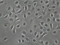
SCC469 MOC1 Mouse Oral Squamous Cell Carcinoma (OSCC) Cell Line

SCC469
≥1X10⁶ cells/vial   
Purchase on Sigma-Aldrich

Overview

Replacement Information
Description
Catalogue NumberSCC469
DescriptionMOC1 Mouse Oral Squamous Cell Carcinoma (OSCC) Cell Line
Alternate Names
  • MOC1 cell line
  • mouse oral carcinoma 1 cell line
  • oral carcinoma cell line
Background InformationDespite our efforts in recent decades to understand growth and signaling pathways in head and neck squamous cell carcinoma (HNSCC), patient outcomes have not improved significantly. A major barrier to understanding and studying immune responses in the context of cancer in recent years has been the lack of available syngeneic mouse models. Xenograft approaches have historically been popular models, but they unfortunately do not provide the critical adaptive immunity components.

The MOC1 cell line, which has a less aggressive growth pattern, generates a tumor microenvironment with higher CD8+ T-cell levels when compared with MOC2. This seems to mirror data that associates a higher CD8+ T-cell count with more favorable outcomes in HNSCC patients.​

The MOC cell lines have expanded our understanding of CXCL14 downregulation in metastatic lymph nodes. Previously, CXCL14 expression had been associated with T-cell associated mechanisms for tumor suppression. Tumor Infiltrating Lymphocyte (TIL) studies using MOC1 and MOC2 have found a cell-specific association in Oral Squamous Cell Carcinomas (OSCCs) involving CXCL14 downregulation resulting in TIL increase which leads to tumor suppression.

The MOC group of cell lines contained common HNSCC mutations in addition to human-related OSCC driver pathways. All MOC lines share RAS pathway mutations and MOC22 was found to have HRAS mutations which is relevant in relation to human HRAS-mutant OSCC groups.

Source:
The MOC1 cell line was derived from carcinogen-induced oral tumors in immunocompetent C57BL/6 mice.

References:
1. Judd, N. P., Allen, C. T., Winkler, A. E., & Uppaluri, R. (2012). Comparative analysis of tumor-infiltrating lymphocytes in a syngeneic mouse model of oral cancer. Otolaryngology--Head and Neck Surgery, 147(3), 493-500.
2. Parikh, A., Shin, J., Faquin, W., Lin, D. T., Tirosh, I., Sunwoo, J. B., & Puram, S. V. (2020). Malignant cell-specific CXCL14 promotes tumor lymphocyte infiltration in oral cavity squamous cell carcinoma. Journal for ImmunoTherapy of Cancer, 8(2).​
3. Onken, M. D., Winkler, A. E., Kanchi, K. L., Chalivendra, V., Law, J. H., Rickert, C. G., ... & Uppaluri, R. (2014). A surprising cross-species conservation in the genomic landscape of mouse and human oral cancer identifies a transcriptional signature predicting metastatic disease. Clinical Cancer Research, 20(11), 2873-2884.
References
Product Information
Components
  • ≥1x10⁶ viable MOC1 Cells: (Catalog No. SCC469). Store in liquid nitrogen.
Quality LevelMQ100
Applications
ApplicationThe MOC (Mouse Oral Cancer) cell lines provide a syngeneic HNSCC cancer model that can be transplanted into C57BL/6 mice. These cell lines are important models for immune cell infiltration applications. The MOC lines have been utilized to compare and identify potential clinical scenarios that are applicable to human Oral Squamous Cell Carcinoma. The MOC1 cell line was derived from primary tumors in a C57BL/6 WT mouse and shows indolent growth compared with the aggressive, metastatic MOC2 line.
Key Applications
  • Cell Culture
Application NotesThis product is intended for sale and sold solely to academic institutions for internal academic research use per the terms of the “Academic Use Agreement” as detailed in the product documentation. For information regarding any other use, please contact licensing@milliporesigma.com.
Biological Information
Cell Line Type
  • Cancer Cells
Physicochemical Information
Dimensions
Materials Information
Toxicological Information
Safety Information according to GHS
Safety Information
Product Usage Statements
Quality Assurance• Each vial contains ≥1X10⁶ viable cells.
• MOC1 cells are verified to be of mouse origin and negative for human, rat, Chinese hamster, Golden Syrian hamster, and non-human primate interspecies contamination, as assessed by a Contamination Clear panel by Charles River Animal Diagnostic Services
• Cells tested negative for infectious diseases against a Mouse Essential CLEAR panel by Charles River Animal Diagnostic Services.
• Cells tested negative for mycoplasma.
Usage Statement
  • Unless otherwise stated in our catalog or other company documentation accompanying the product(s), our products are intended for research use only and are not to be used for any other purpose, which includes but is not limited to, unauthorized commercial uses, in vitro diagnostic uses, ex vivo or in vivo therapeutic uses or any type of consumption or application to humans or animals.
Storage and Shipping Information
Storage ConditionsMOC1 cells should be stored in liquid nitrogen until use. The cells can be cultured for at least 10 passages after initial thawing without significantly affecting cell marker expression and functionality.
Packaging Information
Material Size≥1X10⁶ cells/vial
Transport Information
Supplemental Information
Specifications
Global Trade Item Number
Catalog Number GTIN
SCC469 04065270002396