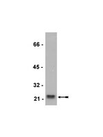
p16 Stimulates CDC42-dependent migration of hepatocellular carcinoma cells.
Chen, YW; Chu, HC; Ze-Shiang Lin, ; Shiah, WJ; Chou, CP; Klimstra, DS; Lewis, BC
PloS one
8
e69389
2013
Show Abstract
Hepatocellular carcinoma (HCC) is a leading cause of cancer-related deaths worldwide. Tumor dissemination to the extra-hepatic region of the portal vein, lymph nodes, lungs or bones contributes to the high mortality seen in HCC; yet, the molecular mechanisms responsible for HCC metastasis remain unclear. Prior studies have suggested a potential link between accumulated cytoplasm-localized p16 and tumor progression. Here we report that p16 enhances metastasis-associated phenotypes in HCC cells - ectopic p16 expression increased cell migration in vitro, and lung colonization after intravenous injection, whereas knockdown of endogenous p16 reduced cell migration. Interestingly, analysis of p16 mutants indicated that the Cdk4 interaction domain is required for stimulation of HCC cell migration; however, knockdown of Cdk4 and Cdk6 showed that these proteins are dispensable for this phenomenon. Intriguingly, we found that in p16-positive HCC samples, p16 protein is predominantly localized in the cytoplasm. In addition, we identified a potential role for nuclear-cytoplasmic shuttling in p16-stimulated migration, consistent with the predominantly cytoplasmic localization of p16 in IHC-positive HCC samples. Finally, we determined that p16-stimulated cell migration requires the Cdc42 GTPase. Our results demonstrate for the first time a pro-migratory role for p16, and suggest a potential mechanism for the observed association between cytoplasmic p16 and tumor progression in diverse tumor types. | | 23894465
 |
Unconjugated Bilirubin Restricts Oligodendrocyte Differentiation and Axonal Myelination.
Barateiro, Andreia, et al.
Mol. Neurobiol., (2012)
2012
Show Abstract
High levels of serum unconjugated bilirubin (UCB) in newborns are associated with axonal damage and glial reactivity that may contribute to subsequent neurologic injury and encephalopathy (kernicterus). Impairments in myelination and white matter damage were observed at autopsy in kernicteric infants. We have recently reported that UCB reduces oligodendrocyte progenitor cell (OPC) survival in a pure OPC in vitro proliferative culture. Here, we hypothesized that neonatal hyperbilirubinemia may also impair oligodendrocyte (OL) maturation and myelination. We used an experimental model of hyperbilirubinemia that has been shown to mimic the pathophysiological conditions leading to brain dysfunction by unbound (free) UCB. Using primary cultures of OL, we demonstrated that UCB delays cell differentiation by increasing the OPC number and reducing the number of mature OL. This finding was combined with a downregulation of Olig1 mRNA levels and upregulation of Olig2 mRNA levels. Addition of UCB, prior to or during differentiation, impaired OL morphological maturation, extension of processes and cell diameter. Both conditions reduced active guanosine triphosphate (GTP)-bound Rac1 fraction. In myelinating co-cultures of dorsal root ganglia neurons and OL, UCB treatment prior to the onset of myelination decreased oligodendroglial differentiation and the number of myelinating OL, also observed when UCB was added after the onset of myelination. In both circumstances, UCB decreased the number of myelin internodes per OL, as well as the myelin internode length. Our studies demonstrate that increased concentrations of UCB compromise myelinogenesis, thereby elucidating a potential deleterious consequence of elevated UCB. | | 23086523
 |
Keratin 8/18 regulation of cell stiffness-extracellular matrix interplay through modulation of Rho-mediated actin cytoskeleton dynamics.
Bordeleau, F; Myrand Lapierre, ME; Sheng, Y; Marceau, N
PloS one
7
e38780
2012
Show Abstract
Cell mechanical activity generated from the interplay between the extracellular matrix (ECM) and the actin cytoskeleton is essential for the regulation of cell adhesion, spreading and migration during normal and cancer development. Keratins are the intermediate filament (IF) proteins of epithelial cells, expressed as pairs in a lineage/differentiation manner. Hepatic epithelial cell IFs are made solely of keratins 8/18 (K8/K18), hallmarks of all simple epithelia. Notably, our recent work on these epithelial cells has revealed a key regulatory function for K8/K18 IFs in adhesion/migration, through modulation of integrin interactions with ECM, actin adaptors and signaling molecules at focal adhesions. Here, using K8-knockdown rat H4 hepatoma cells and their K8/K18-containing counterparts seeded on fibronectin-coated substrata of different rigidities, we show that the K8/K18 IF-lacking cells lose their ability to spread and exhibit an altered actin fiber organization, upon seeding on a low-rigidity substratum. We also demonstrate a concomitant reduction in local cell stiffness at focal adhesions generated by fibronectin-coated microbeads attached to the dorsal cell surface. In addition, we find that this K8/K18 IF modulation of cell stiffness and actin fiber organization occurs through RhoA-ROCK signaling. Together, the results uncover a K8/K18 IF contribution to the cell stiffness-ECM rigidity interplay through a modulation of Rho-dependent actin organization and dynamics in simple epithelial cells. | | 22685604
 |
p53-mediated transcriptional regulation and activation of the actin cytoskeleton regulatory RhoC to LIMK2 signaling pathway promotes cell survival.
Croft, DR; Crighton, D; Samuel, MS; Lourenco, FC; Munro, J; Wood, J; Bensaad, K; Vousden, KH; Sansom, OJ; Ryan, KM; Olson, MF
Cell research
21
666-82
2011
Show Abstract
The central arbiter of cell fate in response to DNA damage is p53, which regulates the expression of genes involved in cell cycle arrest, survival and apoptosis. Although many responses initiated by DNA damage have been characterized, the role of actin cytoskeleton regulators is largely unknown. We now show that RhoC and LIM kinase 2 (LIMK2) are direct p53 target genes induced by genotoxic agents. Although RhoC and LIMK2 have well-established roles in actin cytoskeleton regulation, our results indicate that activation of LIMK2 also has a pro-survival function following DNA damage. LIMK inhibition by siRNA-mediated knockdown or selective pharmacological blockade sensitized cells to radio- or chemotherapy, such that treatments that were sub-lethal when administered singly resulted in cell death when combined with LIMK inhibition. Our findings suggest that combining LIMK inhibitors with genotoxic therapies could be more efficacious than single-agent administration, and highlight a novel connection between actin cytoskeleton regulators and DNA damage-induced cell survival mechanisms. | | 21079653
 |
Early intervention of tyrosine nitration prevents vaso-obliteration and neovascularization in ischemic retinopathy.
Abdelsaid, Mohammed A, et al.
J. Pharmacol. Exp. Ther., 332: 125-34 (2010)
2010
Show Abstract
Diabetic retinopathy and retinopathy of prematurity are blinding disorders that follow a pathological pattern of ischemic retinopathy and affect premature infants and working-age adults. Yet, the treatment options are limited to laser photocoagulation. The goal of this study is to elucidate the molecular mechanism and examine the therapeutic effects of inhibiting tyrosine nitration on protecting early retinal vascular cell death and late neovascularization in the ischemic retinopathy model. Ischemic retinopathy was developed by exposing neonatal mice to 75% oxygen [postnatal day (p) 7-p12] followed by normoxia (21% oxygen) (p12-p17). Peroxynitrite decomposition catalyst 5,10,15,20-tetrakis(4-sulfonatophenyl)porphyrinato iron III chloride (FeTPPS) (1 mg/kg), the nitration inhibitor epicatechin (10 mg/kg) or the thiol donor N-acetylcysteine (NAC, 150 mg/kg) were administered (p7-p12) or (p7-p17). Vascular endothelial cells were incubated at hyperoxia (40% oxygen) or normoxia (21% oxygen) for 48 h. Vascular density was determined in retinal flat mounts labeled with isolectin B4. Expression of vascular endothelial growth factor, caspase-3, and poly(ADP ribose) polymerase (PARP), activation of Akt and p38 mitogen-activated protein kinase (MAPK), and tyrosine nitration of the phosphatidylinositol (PI) 3-kinase p85 subunit were analyzed by Western blot. Hyperoxia-induced peroxynitrite caused endothelial cell apoptosis as indicated by expression of cleaved caspase-3 and PARP leading to vaso-obliteration. These effects were associated with significant tyrosine nitration of the p85 subunit of PI 3-kinase, decreased Akt activation, and enhanced p38 MAPK activation. Blocking tyrosine nitration of PI 3-kinase with epicatechin or NAC restored Akt phosphorylation, and inhibited vaso-obliteration at p12 and neovascularization at p17 comparable with FeTPPS. Early inhibition of tyrosine nitration with use of epicatechin or NAC can represent safe and effective vascular-protective agents in ischemic retinopathy. | | 19815813
 |
Peroxynitrite mediates diabetes-induced endothelial dysfunction: possible role of Rho kinase activation.
El-Remessy, Azza B, et al.
Exp Diabetes Res, 2010: 247861 (2010)
2010
Show Abstract
Endothelial dysfunction is characterized by reduced bioavailability of NO due to its inactivation to form peroxynitrite or reduced expression of eNOS. Here, we examine the causal role of peroxynitrite in mediating diabetes-induced endothelial dysfunction. Diabetes was induced by STZ-injection, and rats received the peroxynitrite decomposition catalyst (FeTTPs, 15 mg/Kg/day) for 4 weeks. Vasorelaxation to acetylcholine, oxidative-stress markers, RhoA activity, and eNOS expression were determined. Diabetic coronary arteries showed significant reduction in ACh-mediated maximal relaxation compared to controls. Diabetic vessels showed also significant increases in lipid-peroxides, nitrotyrosine, and active RhoA and 50% reduction in eNOS mRNA expression. Treatment of diabetic animals with FeTTPS blocked these effects. Studies in aortic endothelial cells show that high glucose or peroxynitrite increases the active RhoA kinase levels and decreases eNOS expression and NO levels, which were reversed with blocking peroxynitrite or Rho kinase. Together, peroxynitrite can suppress eNOS expression via activation of RhoA and hence cause vascular dysfunction. | | 21052489
 |
Ascorbate-induced osteoblast differentiation recruits distinct MMP-inhibitors: RECK and TIMP-2.
Willian F Zambuzzi,Claudia L Yano,Alexandre D M Cavagis,Maikel P Peppelenbosch,José Mauro Granjeiro,Carmen V Ferreira
Molecular and cellular biochemistry
322
2009
Show Abstract
The bone formation executed by osteoblasts represents an interesting research field both for basic and applied investigations. The goal of this work was to evaluate the molecular mechanisms involved during osteoblast differentiation in vitro. Accordingly, we demonstrated that, during the osteoblastic differentiation, TIMP-2 and RECK presented differential expressions, where RECK expression was downregulated from the 14th day in contrast with an increase in TIMP-2. Concomitantly, our results showed a temporal regulation of two major signaling cascades during osteoblast differentiation: proliferation cascades in which RECK, PI3 K, and GSK-3beta play a pivotal role and latter, differentiation cascades with participation of Ras, Rho, Rac-1, PKC alpha/beta, and TIMP-2. Furthermore, we observed that phosphorylation level of paxillin was downregulated while FAK(125) remained unchangeable, but active during extracellular matrix (ECM) remodeling. Concluding, our results provide evidences that RECK and TIMP-2 are involved in the control of ECM remodeling in distinct phases of osteoblast differentiation by modulating MMP activities and a multitude of signaling proteins governs these events. | | 18989628
 |
Cdk5-dependent regulation of Rho activity, cytoskeletal contraction, and epithelial cell migration via suppression of Src and p190RhoGAP.
Tripathi, BK; Zelenka, PS
Molecular and cellular biology
29
6488-99
2009
Show Abstract
Cdk5 regulates adhesion and migration in a variety of cell types. We previously showed that Cdk5 is strongly activated during stress fiber formation and contraction in spreading cells. Here we determine the mechanism linking Cdk5 to stress fiber contractility and its relevance to cell migration. Immunofluorescence showed that Cdk5 colocalized with phosphorylated myosin regulatory light chain (pMRLC) on contracting stress fibers. Inhibiting Cdk5 activity by various means significantly reduced pMRLC level and cytoskeletal contraction, with loss of central stress fibers. Blocking Cdk5 activity also reduced Rho-Rho kinase (ROCK) signaling, which is the principal pathway of myosin phosphorylation under these conditions. Next, we examined the effect of Cdk5 activity on Src, a known regulator of Rho. Inhibiting Cdk5 activity increased Src activation and phosphorylation of its substrate, p190RhoGAP, an upstream inhibitor of Rho. Inhibiting both Cdk5 and Src activity completely reversed the effect of Cdk5 inhibition on Rho and prevented the loss of central stress fibers, demonstrating that Cdk5 exerts its effects on Rho-ROCK signaling by suppressing Src activity. Moreover, inhibiting either Cdk5 or ROCK activity increased cell migration to an equal extent, while inhibiting both kinases produced no additional effect, demonstrating that Cdk5-dependent regulation of ROCK activity is a physiological determinant of migration rate. Full Text Article | | 19822667
 |
Atorvastatin inhibits ABCA1 expression and cholesterol efflux in THP-1 macrophages by an LXR-dependent pathway.
Guosong Qiu,John S Hill
Journal of cardiovascular pharmacology
51
2008
Show Abstract
The effect of atorvastatin on adenosine triphosphate (ATP)-binding cassette transporter A1 (ABCA1) expression and cholesterol efflux remains controversial. In an effort to clarify this issue, ABCA1 expression and apolipoprotein AI (apoAI)-mediated cholesterol efflux after atorvastatin treatment were investigated in THP-1 macrophages. Atorvastatin from 2 microM to 40 microM dose-dependently inhibited ABCA1 expression in human monocyte-derived macrophages and phorbol 12-myristate 13-acetate (PMA)-stimulated THP-1 monocytes. ApoAI-mediated cholesterol efflux was reduced in PMA-stimulated THP-1 cells treated with atorvastatin, this effect was abolished with acetylated low-density lipoprotein (LDL) pretreatment. Atorvastatin treatment also dose-dependently reduced liver X receptor alpha (LXRalpha) expression and Rho activation. Rho activation by farnysylpyophosphate (FPP) and lysophosphatidic acid (LPA) did not salvage, but further depressed, the cholesterol efflux and ABCA1 expression in the presence of atorvastatin. Without atorvastatin, Rho activation by mevalonate, FPP, and LPA diminished apoAI-mediated cholesterol efflux, and Rho activation by GTPgammaS also decreased ABCA1 messenger ribonucleic acid (mRNA) by 16%. Furthermore, Rho inhibition by C3 exoenzyme increased ABCA1 mRNA by 48% despite a 17% decrease in apoAI-mediated cholesterol efflux. LXRalpha agonists (T01901317 and 22(R)-hydroxycholesterol) prevented any reductions in cholesterol efflux or ABCA1 expression associated with atorvastatin treatment. Furthermore, Western blot analysis demonstrated the reciprocal inhibition of Rho and LXRalpha. In conclusion, atorvastatin decreases ABCA1 expression in noncholesterol-loaded macrophages in an LXRalpha- but not Rho-dependent pathway; this effect can be compromised after acetylated LDL cholesterol loading. | | 18427282
 |
G alpha o mediates WNT-JNK signaling through dishevelled 1 and 3, RhoA family members, and MEKK 1 and 4 in mammalian cells.
Bikkavilli, RK; Feigin, ME; Malbon, CC
Journal of cell science
121
234-45
2008
Show Abstract
In Drosophila, activation of Jun N-terminal Kinase (JNK) mediated by Frizzled and Dishevelled leads to signaling linked to planar cell polarity. A biochemical delineation of WNT-JNK planar cell polarity was sought in mammalian cells, making use of totipotent mouse F9 teratocarcinoma cells that respond to WNT3a via Frizzled-1. The canonical WNT-beta-catenin signaling pathway requires both G alpha o and G alpha q heterotrimeric G-proteins, whereas we show that WNT-JNK signaling requires only G alpha o protein. G alpha o propagates the signal downstream through all three Dishevelled isoforms, as determined by epistasis experiments using the Dishevelled antagonist Dapper1 (DACT1). Suppression of either Dishevelled-1 or Dishevelled-3, but not Dishevelled-2, abolishes WNT3a activation of JNK. Activation of the small GTPases RhoA, Rac1 and Cdc42 operates downstream of Dishevelled, linking to the MEKK 1/MEKK 4-dependent cascade, and on to JNK activation. Chemical inhibitors of JNK (SP600125), but not p38 (SB203580), block WNT3a activation of JNK, whereas both the inhibitors attenuate the WNT3a-beta-catenin pathway. These data reveal both common and unique signaling elements in WNT3a-sensitive pathways, highlighting crosstalk from WNT3a-JNK to WNT3a-beta-catenin signaling. | Western Blotting | 18187455
 |


