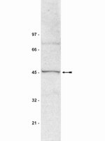
Suv39h1 mediates AP-2α-dependent inhibition of C/EBPα expression during adipogenesis.
Zhang, ZC; Liu, Y; Li, SF; Guo, L; Zhao, Y; Qian, SW; Wen, B; Tang, QQ; Li, X
Molecular and cellular biology
34
2330-8
2014
Show Abstract
Previous studies have shown that CCAAT/enhancer-binding protein α (C/EBPα) plays a very important role during adipocyte terminal differentiation and that AP-2α (activator protein 2α) acts as a repressor to delay the expression of C/EBPα. However, the mechanisms by which AP-2α prevents the expression of C/EBPα are not fully understood. Here, we present evidence that Suv39h1, a histone H3 lysine 9 (H3K9)-specific trimethyltransferase, and G9a, a euchromatic methyltransferase, both interact with AP-2α and enhance AP-2α-mediated transcriptional repression of C/EBPα. Interestingly, we discovered that G9a mediates dimethylation of H3K9, thus providing the substrate, which is methylated by Suv39h1, to H3K9me3 on the C/EBPα promoter. The expression level of AP-2α was consistent with enrichment of H3K9me2 and H3K9me3 on the C/EBPα promoter in 3T3-L1 preadipocytes. Knockdown of Suv39h markedly increased C/EBPα expression and promoted adipogenesis. Conversely, ectopic expression of Suv39h1 delayed C/EBPα expression and impaired the accumulation of triglyceride, while simultaneous knockdown of AP-2α or G9a partially rescued this process. These findings indicate that Suv39h1 enhances AP-2α-mediated transcriptional repression of C/EBPα in an epigenetic manner and further inhibits adipocyte differentiation. | Western Blotting | 24732798
 |
SPOC1 modulates DNA repair by regulating key determinants of chromatin compaction and DNA damage response.
Mund, A; Schubert, T; Staege, H; Kinkley, S; Reumann, K; Kriegs, M; Fritsch, L; Battisti, V; Ait-Si-Ali, S; Hoffbeck, AS; Soutoglou, E; Will, H
Nucleic acids research
40
11363-79
2012
Show Abstract
Survival time-associated plant homeodomain (PHD) finger protein in Ovarian Cancer 1 (SPOC1, also known as PHF13) is known to modulate chromatin structure and is essential for testicular stem-cell differentiation. Here we show that SPOC1 is recruited to DNA double-strand breaks (DSBs) in an ATM-dependent manner. Moreover, SPOC1 localizes at endogenous repair foci, including OPT domains and accumulates at large DSB repair foci characteristic for delayed repair at heterochromatic sites. SPOC1 depletion enhances the kinetics of ionizing radiation-induced foci (IRIF) formation after γ-irradiation (γ-IR), non-homologous end-joining (NHEJ) repair activity, and cellular radioresistance, but impairs homologous recombination (HR) repair. Conversely, SPOC1 overexpression delays IRIF formation and γH2AX expansion, reduces NHEJ repair activity and enhances cellular radiosensitivity. SPOC1 mediates dose-dependent changes in chromatin association of DNA compaction factors KAP-1, HP1-α and H3K9 methyltransferases (KMT) GLP, G9A and SETDB1. In addition, SPOC1 interacts with KAP-1 and H3K9 KMTs, inhibits KAP-1 phosphorylation and enhances H3K9 trimethylation. These findings provide the first evidence for a function of SPOC1 in DNA damage response (DDR) and repair. SPOC1 acts as a modulator of repair kinetics and choice of pathways. This involves its dose-dependent effects on DNA damage sensors, repair mediators and key regulators of chromatin structure. | | 23034801
 |
5-Aza-2'-deoxycytidine reactivates gene expression via degradation of pRb pocket proteins.
Zheng, Z; Li, L; Liu, X; Wang, D; Tu, B; Wang, L; Wang, H; Zhu, WG
FASEB journal : official publication of the Federation of American Societies for Experimental Biology
26
449-59
2012
Show Abstract
Not only does 5-aza-2'-deoxycytidine (5-aza-CdR) induce the reexpression of silenced genes through the demethylation of CpG islands, but it increases the expression of unmethylated genes. However, the mechanism by which 5-aza-CdR activates the expression of genes is not completely understood. Here, we report that the pRb pocket proteins pRb, p107, and p130 were degraded in various cancer cell lines in response to 5-aza-CdR treatment, and this effect was dependent on the proteasome pathway. Mouse double minute 2 (MDM2) played a critical role in this 5-aza-CdR-induced degradation of pRb. Furthermore, PP2A phosphatase-induced MDM2 dephosphorylation at S260 was found to be essential for MDM2 binding to pRb in the presence of 5-aza-CdR. pRb degradation resulted in the significant reexpression of several genes, including methylated CDKN2A, RASFF1A, and unmethylated CDKN2D. Finally, knockdown of pRb pocket proteins by either RNAi or 5-aza-CdR treatment induced a significant decrease in the recruitment of SUV39H1 and an increase in the enrichment of KDM3B and KDM4A to histones around the promoter of RASFF1A and thus reduced H3K9 di- and trimethylation, by which RASFF1A expression is activated. Our data reveal a novel mechanism by which 5-aza-CdR induces the expression of both methylated and unmethylated genes by degrading pRb pocket proteins. | | 21990374
 |
p53-mediated heterochromatin reorganization regulates its cell fate decisions.
Mungamuri, SK; Benson, EK; Wang, S; Gu, W; Lee, SW; Aaronson, SA
Nature structural & molecular biology
19
478-84, S1
2012
Show Abstract
p53 is a major sensor of cellular stresses, and its activation influences cell fate decisions. We identified SUV39H1, a histone code 'writer' responsible for the histone H3 Lys9 trimethylation (H3K9me3) mark for 'closed' chromatin conformation, as a target of p53 repression. SUV39H1 downregulation was mediated transcriptionally by p21 and post-translationally by MDM2. The H3K9me3 repression mark was found to be associated with promoters of representative p53 target genes and was decreased upon p53 activation. Overexpression of SUV39H1 maintained higher levels of the H3K9me3 mark on these promoters and was associated with decreased p53 promoter occupancy and decreased transcriptional induction in response to p53. Conversely, SUV39H1 pre-silencing decreased H3K9me3 levels on these promoters and enhanced the p53 apoptotic response. These findings uncover a new layer of p53-mediated chromatin regulation through modulation of histone methylation at p53 target promoters. | Western Blotting | 22466965
 |
Adenomatous polyposis coli and hypoxia-inducible factor-1{alpha} have an antagonistic connection.
Newton, IP; Kenneth, NS; Appleton, PL; Näthke, I; Rocha, S
Molecular biology of the cell
21
3630-8
2010
Show Abstract
The tumor suppressor adenomatous polyposis coli (APC) is mutated in the majority of colorectal cancers and is best known for its role as a scaffold in a Wnt-regulated protein complex that determines the availability of β-catenin. Another common feature of solid tumors is the presence of hypoxia as indicated by the up-regulation of hypoxia-inducible factors (HIFs) such as HIF-1α. Here, we demonstrate a novel link between APC and hypoxia and show that APC and HIF-1α antagonize each other. Hypoxia results in reduced levels of APC mRNA and protein via a HIF-1α-dependent mechanism. HIF-1α represses the APC gene via a functional hypoxia-responsive element on the APC promoter. In contrast, APC-mediated repression of HIF-1α requires wild-type APC, low levels of β-catenin, and nuclear factor-κB activity. These results reveal down-regulation of APC as a new mechanism that contributes to the survival advantage induced by hypoxia and also show that loss of APC mutations produces a survival advantage by mimicking hypoxic conditions. Full Text Article | | 20844082
 |
Enhanced levels of microRNA-125b in vascular smooth muscle cells of diabetic db/db mice lead to increased inflammatory gene expression by targeting the histone methyltransferase Suv39h1.
Villeneuve, LM; Kato, M; Reddy, MA; Wang, M; Lanting, L; Natarajan, R
Diabetes
59
2904-15
2010
Show Abstract
Diabetes remains a major risk factor for vascular complications that seem to persist even after achieving glycemic control, possibly due to "metabolic memory." Using cultured vascular smooth muscle cells (MVSMC) from type 2 diabetic db/db mice, we recently showed that decreased promoter occupancy of the chromatin histone H3 lysine-9 methyltransferase Suv39h1 and the associated repressive epigenetic mark histone H3 lysine-9 trimethylation (H3K9me3) play key roles in sustained inflammatory gene expression. Here we examined the role of microRNAs (miRs) in Suv39h1 regulation and function in MVSMC from diabetic mice.We used luciferase assays with Suv39h1 3'untranslated region (UTR) reporter constructs and Western blotting of endogenous protein to verify that miR-125b targets Suv39h1. We examined the effects of Suv39h1 targeting on inflammatory gene expression by quantitative real time polymerase chain reaction (RT-qPCR), and H3K9me3 levels at their promoters by chromatin immunoprecipitation assays.We observed significant upregulation of miR-125b with parallel downregulation of Suv39h1 protein (predicted miR-125b target) in MVSMC cultured from diabetic db/db mice relative to control db/+. miR-125b mimics inhibited both Suv39h1 3'UTR luciferase reporter activity and endogenous Suv39h1 protein levels. Conversely, miR-125b inhibitors showed opposite effects. Furthermore, miR-125b mimics increased expression of inflammatory genes, monocyte chemoattractant protein-1, and interleukin-6, and reduced H3K9me3 at their promoters in nondiabetic cells. Interestingly, miR-125b mimics increased monocyte binding to db/+ MVSMC toward that in db/db MVSMC, further imitating the proinflammatory diabetic phenotype. In addition, we found that the increase in miR-125b in db/db VSMC is caused by increased transcription of miR-125b-2.These results demonstrate a novel upstream role for miR-125b in the epigenetic regulation of inflammatory genes in MVSMC of db/db mice through downregulation of Suv39h1. Full Text Article | | 20699419
 |
ASXL1 represses retinoic acid receptor-mediated transcription through associating with HP1 and LSD1.
Lee SW, Cho YS, Na JM, Park UH, Kang M, Kim EJ, Um SJ
The Journal of biological chemistry
285
18-29
2010
Show Abstract
We previously suggested that ASXL1 (additional sex comb-like 1) functions as either a coactivator or corepressor for the retinoid receptors retinoic acid receptor (RAR) and retinoid X receptor in a cell type-specific manner. Here, we provide clues toward the mechanism underlying ASXL1-mediated repression. Transfection assays in HEK293 or H1299 cells indicated that ASXL1 alone possessing autonomous transcriptional repression activity significantly represses RAR- or retinoid X receptor-dependent transcriptional activation, and the N-terminal portion of ASXL1 is responsible for the repression. Amino acid sequence analysis identified a consensus HP1 (heterochromatin protein 1)-binding site (HP1 box, PXVXL) in that region. Systematic in vitro and in vivo assays revealed that the HP1 box in ASXL1 is critical for the interaction with the chromoshadow domain of HP1. Transcription assays with HP1 box deletion or HP1alpha knockdown indicated that HP1alpha is required for ASXL1-mediated repression. Furthermore, we found a direct interaction of ASXL1 with histone H3 demethylase LSD1 through the N-terminal region nearby the HP1-binding site. ASXL1 binding to LSD1 was greatly increased by HP1alpha, resulting in the formation of a ternary complex. LSD1 cooperates with ASXL1 in transcriptional repression, presumably by removing H3K4 methylation, an active histone mark, but not H3K9 methylation, a repressive histone mark recognized by HP1. This possibility was supported by chromatin immunoprecipitation assays followed by ASXL1 overexpression or knockdown. Overall, this study provides the first evidence that ASXL1 cooperates with HP1 to modulate LSD1 activity, leading to a change in histone H3 methylation and thereby RAR repression. Full Text Article | | 19880879
 |
Epidermal growth factor receptor regulates beta-catenin location, stability, and transcriptional activity in oral cancer.
Lee, CH; Hung, HW; Hung, PH; Shieh, YS
Molecular cancer
9
64
2010
Show Abstract
Many cancerous cells accumulate beta-catenin in the nucleus. We examined the role of epidermal growth factor receptor (EGFR) signaling in the accumulation of beta-catenin in the nuclei of oral cancer cells.We used two strains of cultured oral cancer cells, one with reduced EGFR expression (OECM1 cells) and one with elevated EGFR expression (SAS cells), and measured downstream effects, such as phosphorylation of beta-catenin and GSK-3beta, association of beta-catenin with E-cadherin, and target gene regulation. We also studied the expression of EGFR, beta-catenin, and cyclin D1 in 112 samples of oral cancer by immunostaining. Activation of EGFR signaling increased the amount of beta-catenin in the nucleus and decreased the amount in the membranes. EGF treatment increased phosphorylation of beta-catenin (tyrosine) and GSK-3beta(Ser-(9), resulting in a loss of beta-catenin association with E-cadherin. TOP-FLASH and FOP-FLASH reporter assays demonstrated that the EGFR signal regulates beta-catenin transcriptional activity and mediates cyclin D1 expression. Chromatin immunoprecipitation experiments indicated that the EGFR signal affects chromatin architecture at the regulatory element of cyclin D1, and that the CBP, HDAC1, and Suv39h1 histone/chromatin remodeling complex is involved in this process. Immunostaining showed a significant association between EGFR expression and aberrant accumulation of beta-catenin in oral cancer.EGFR signaling regulates beta-catenin localization and stability, target gene expression, and tumor progression in oral cancer. Moreover, our data suggest that aberrant accumulation of beta-catenin under EGFR activation is a malignancy marker of oral cancer. | | 20302655
 |
Hyperglycemia induces a dynamic cooperativity of histone methylase and demethylase enzymes associated with gene-activating epigenetic marks that coexist on the lysine tail
Daniella Brasacchio 1 , Jun Okabe, Christos Tikellis, Aneta Balcerczyk, Prince George, Emma K Baker, Anna C Calkin, Michael Brownlee, Mark E Cooper, Assam El-Osta
Diabetes
58(5)
1229-36
2009
Show Abstract
Objective: Results from the Diabetes Control Complications Trial (DCCT) and the subsequent Epidemiology of Diabetes Interventions and Complications (EDIC) Study and more recently from the U.K. Prospective Diabetes Study (UKPDS) have revealed that the deleterious end-organ effects that occurred in both conventional and more aggressively treated subjects continued to operate >5 years after the patients had returned to usual glycemic control and is interpreted as a legacy of past glycemia known as "hyperglycemic memory." We have hypothesized that transient hyperglycemia mediates persistent gene-activating events attributed to changes in epigenetic information. <br />Research design and methods: Models of transient hyperglycemia were used to link NFkappaB-p65 gene expression with H3K4 and H3K9 modifications mediated by the histone methyltransferases (Set7 and SuV39h1) and the lysine-specific demethylase (LSD1) by the immunopurification of soluble NFkappaB-p65 chromatin. <br />Results: The sustained upregulation of the NFkappaB-p65 gene as a result of ambient or prior hyperglycemia was associated with increased H3K4m1 but not H3K4m2 or H3K4m3. Furthermore, glucose was shown to have other epigenetic effects, including the suppression of H3K9m2 and H3K9m3 methylation on the p65 promoter. Finally, there was increased recruitment of the recently identified histone demethylase LSD1 to the p65 promoter as a result of prior hyperglycemia. <br />Conclusions: These studies indicate that the active transcriptional state of the NFkappaB-p65 gene is linked with persisting epigenetic marks such as enhanced H3K4 and reduced H3K9 methylation, which appear to occur as a result of effects of the methyl-writing and methyl-erasing histone enzymes. | | 19208907
 |
Histone deacetylase inhibitor depsipeptide activates silenced genes through decreasing both CpG and H3K9 methylation on the promoter.
Wu, LP; Wang, X; Li, L; Zhao, Y; Lu, S; Yu, Y; Zhou, W; Liu, X; Yang, J; Zheng, Z; Zhang, H; Feng, J; Yang, Y; Wang, H; Zhu, WG
Molecular and cellular biology
28
3219-35
2008
Show Abstract
Histone deacetylase inhibitor (HDACi) has been shown to demethylate the mammalian genome, which further strengthens the concept that DNA methylation and histone modifications interact in regulation of gene expression. Here, we report that an HDAC inhibitor, depsipeptide, exhibited significant demethylating activity on the promoters of several genes, including p16, SALL3, and GATA4 in human lung cancer cell lines H719 and H23, colon cancer cell line HT-29, and pancreatic cancer cell line PANC1. Although expression of DNA methyltransferase 1 (DNMT1) was not affected by depsipeptide, a decrease in binding of DNMT1 to the promoter of these genes played a dominant role in depsipeptide-induced demethylation and reactivation. Depsipeptide also suppressed expression of histone methyltransferases G9A and SUV39H1, which in turn resulted in a decrease of di- and trimethylated H3K9 around these genes' promoter. Furthermore, both loading of heterochromatin-associated protein 1 (HP1alpha and HP1beta) to methylated H3K9 and binding of DNMT1 to these genes' promoter were significantly reduced in depsipeptide-treated cells. Similar DNA demethylation was induced by another HDAC inhibitor, apicidin, but not by trichostatin A. Our data describe a novel mechanism of HDACi-mediated DNA demethylation via suppression of histone methyltransferases and reduced recruitment of HP1 and DNMT1 to the genes' promoter. Full Text Article | | 18332107
 |


