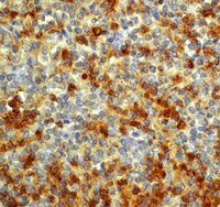
Neuregulin-1 inhibits neuroinflammatory responses in a rat model of organophosphate-nerve agent-induced delayed neuronal injury.
Li, Y; Lein, PJ; Ford, GD; Liu, C; Stovall, KC; White, TE; Bruun, DA; Tewolde, T; Gates, AS; Distel, TJ; Surles-Zeigler, MC; Ford, BD
Journal of neuroinflammation
12
64
2015
Show Abstract
Neuregulin-1 (NRG-1) has been shown to act as a neuroprotectant in animal models of nerve agent intoxication and other acute brain injuries. We recently demonstrated that NRG-1 blocked delayed neuronal death in rats intoxicated with the organophosphate (OP) neurotoxin diisopropylflurophosphate (DFP). It has been proposed that inflammatory mediators are involved in the pathogenesis of OP neurotoxin-mediated brain damage.We examined the influence of NRG-1 on inflammatory responses in the rat brain following DFP intoxication. Microglial activation was determined by immunohistchemistry using anti-CD11b and anti-ED1 antibodies. Gene expression profiling was performed with brain tissues using Affymetrix gene arrays and analyzed using the Ingenuity Pathway Analysis software. Cytokine mRNA levels following DFP and NRG-1 treatment was validated by real-time reverse transcription polymerase chain reaction (RT-PCR).DFP administration resulted in microglial activation in multiple brain regions, and this response was suppressed by treatment with NRG-1. Using microarray gene expression profiling, we observed that DFP increased mRNA levels of approximately 1,300 genes in the hippocampus 24 h after administration. NRG-1 treatment suppressed by 50% or more a small fraction of DFP-induced genes, which were primarily associated with inflammatory responses. Real-time RT-PCR confirmed that the mRNAs for pro-inflammatory cytokines interleukin-1β (IL-1β) and interleukin-6 (IL-6) were significantly increased following DFP exposure and that NRG-1 significantly attenuated this transcriptional response. In contrast, tumor necrosis factor α (TNFα) transcript levels were unchanged in both DFP and DFP + NRG-1 treated brains relative to controls.Neuroprotection by NRG-1 against OP neurotoxicity is associated with the suppression of pro-inflammatory responses in brain microglia. These findings provide new insight regarding the molecular mechanisms involved in the neuroprotective role of NRG-1 in acute brain injuries. | | | 25880399
 |
Feasibility and safety of continuous and chronic bilateral deep brain stimulation of the medial forebrain bundle in the naïve Sprague-Dawley rat.
Furlanetti, LL; Döbrössy, MD; Aranda, IA; Coenen, VA
Behavioural neurology
2015
256196
2015
Show Abstract
Deep brain stimulation (DBS) of the superolateral branch of the medial forebrain bundle (MFB) has provided rapid and dramatic reduction of depressive symptoms in a clinical trial. Early intracranial self-stimulation experiments of the MFB suggested detrimental side effects on the animals' health; therefore, the current study looked at the viability of chronic and continuous MFB-DBS in rodents, with particular attention given to welfare issues and identification of stimulated pathways.Sprague-Dawley female rats were submitted to stereotactic microelectrode implantation into the MFB. Chronic continuous DBS was applied for 3-6 weeks. Welfare monitoring and behavior changes were assessed. Postmortem histological analysis of c-fos protein expression was carried out.MFB-DBS resulted in mild and temporary weight loss in the animals, which was regained even with continuing stimulation. MFB-DBS led to increased and long-lasting c-fos expression in target regions of the mesolimbic/mesocortical system.Bilateral continuous chronic MFB-DBS is feasible, safe, and without impact on the rodent's health. MFB-DBS results in temporary increase in exploration, which could explain the initial weight loss, and does not produce any apparent behavioral abnormalities. This platform represents a powerful tool for further preclinical investigation of the MFB stimulation in the treatment of depression. | | | 25960609
 |
Transient but not permanent benefit of neuronal progenitor cell therapy after traumatic brain injury: potential causes and translational consequences.
Skardelly, M; Gaber, K; Burdack, S; Scheidt, F; Schuhmann, MU; Hilbig, H; Meixensberger, J; Boltze, J
Frontiers in cellular neuroscience
8
318
2014
Show Abstract
Numerous studies have reported a beneficial impact of neural progenitor cell transplantation on functional outcome after traumatic brain injury (TBI) during short and medium follow-up periods. However, our knowledge regarding long-term functional effects is fragmentary while a direct comparison between local and systemic transplantation is missing so far.This study investigated the long-term (12 week) impact of human fetal neuronal progenitor cell (hNPC) transplantation 24 h after severe TBI in rats.Cells were either transplanted stereotactically (1 × 10(5)) into the putamen or systemically (5 × 10(5)) via the tail vein. Control animals received intravenous transplantation of vehicle solution.An overall functional benefit was observed after systemic, but not local hNPC transplantation by area under the curve analysis (p less than 0.01). Surprisingly, this effect vanished during later stages after TBI with all groups exhibiting comparable functional outcomes 84 days after TBI. Investigation of cell-mediated inflammatory processes revealed increasing microglial activation and macrophage presence during these stages, which was statistically significant after systemic cell administration (p less than 0.05). Intracerebral hNPC transplantation slightly diminished astrogliosis in perilesional areas (p less than 0.01), but did not translate into a permanent functional benefit. No significant effects on angiogenesis were observed among the groups.Our results suggest the careful long-term assessment of cell therapies for TBI, as well as to identify potential long-term detrimental effects of such therapies before moving on to clinical trials. Moreover, immunosuppressive protocols, though widely used, should be rigorously assessed for their applicability in the respective setup. | | | 25352780
 |
Distinctive tooth-extraction socket healing: bisphosphonate versus parathyroid hormone therapy.
Kuroshima, S; Mecano, RB; Tanoue, R; Koi, K; Yamashita, J
Journal of periodontology
85
24-33
2014
Show Abstract
Patients with osteoporosis who receive tooth extractions are typically on either oral bisphosphonate or parathyroid hormone (PTH) therapy. Currently, the consequence of these therapies on hard- and soft-tissue healing in the oral cavity is not clearly defined. The aim of this study is to determine the differences in the therapeutic effect on tooth-extraction wound healing between bisphosphonate and PTH therapies.Maxillary second molars were extracted in Sprague Dawley rats (n = 30), and either bisphosphonate (zoledronate [Zol]), PTH, or saline (vehicle control [VC]) was administered for 10 days (n = 10 per group). Hard-tissue healing was evaluated by microcomputed tomography and histomorphometric analyses. Collagen, blood vessels, inflammatory cell infiltration, and cathepsin K expression were assessed in soft tissue using immunohistochemistry, quantitative polymerase chain reaction, and immunoblotting.Both therapies significantly increased bone fill and suppressed vertical bone loss. However, considerably more devital bone was observed in the sockets of rats on Zol versus VC. Although Zol increased the numbers of blood vessels, the total blood vessel area in soft tissue was significantly smaller than in VC. PTH therapy increased osteoblastic bone formation and suppressed osteoclasts. PTH therapy promoted soft-tissue maturation by suppressing inflammation and stimulating collagen deposition.Zoledronate therapy deters whereas PTH therapy promotes hard- and soft-tissue healing in the oral cavity, and both therapies prevent vertical bone loss. | | | 23688101
 |
Abiotic-biotic characterization of Pt/Ir microelectrode arrays in chronic implants.
Prasad, A; Xue, QS; Dieme, R; Sankar, V; Mayrand, RC; Nishida, T; Streit, WJ; Sanchez, JC
Frontiers in neuroengineering
7
2
2014
Show Abstract
Pt/Ir electrodes have been extensively used in neurophysiology research in recent years as they provide a more inert recording surface as compared to tungsten or stainless steel. While floating microelectrode arrays (FMA) consisting of Pt/Ir electrodes are an option for neuroprosthetic applications, long-term in vivo functional performance characterization of these FMAs is lacking. In this study, we have performed comprehensive abiotic-biotic characterization of Pt/Ir arrays in 12 rats with implant periods ranging from 1 week up to 6 months. Each of the FMAs consisted of 16-channel, 1.5 mm long, and 75 μm diameter microwires with tapered tips that were implanted into the somatosensory cortex. Abiotic characterization included (1) pre-implant and post-explant scanning electron microscopy (SEM) to study recording site changes, insulation delamination and cracking, and (2) chronic in vivo electrode impedance spectroscopy. Biotic characterization included study of microglial responses using a panel of antibodies, such as Iba1, ED1, and anti-ferritin, the latter being indicative of blood-brain barrier (BBB) disruption. Significant structural variation was observed pre-implantation among the arrays in the form of irregular insulation, cracks in insulation/recording surface, and insulation delamination. We observed delamination and cracking of insulation in almost all electrodes post-implantation. These changes altered the electrochemical surface area of the electrodes and resulted in declining impedance over the long-term due to formation of electrical leakage pathways. In general, the decline in impedance corresponded with poor electrode functional performance, which was quantified via electrode yield. Our abiotic results suggest that manufacturing variability and insulation material as an important factor contributing to electrode failure. Biotic results show that electrode performance was not correlated with microglial activation (neuroinflammation) as we were able to observe poor performance in the absence of neuroinflammation, as well as good performance in the presence of neuroinflammation. One biotic change that correlated well with poor electrode performance was intraparenchymal bleeding, which was evident macroscopically in some rats and presented microscopically by intense ferritin immunoreactivity in microglia/macrophages. Thus, we currently consider intraparenchymal bleeding, suboptimal electrode fabrication, and insulation delamination as the major factors contributing toward electrode failure. | | | 24550823
 |
Mitigation of diabetes-related complications in implanted collagen and elastin scaffolds using matrix-binding polyphenol.
Chow, JP; Simionescu, DT; Warner, H; Wang, B; Patnaik, SS; Liao, J; Simionescu, A
Biomaterials
34
685-95
2013
Show Abstract
There is a major need for scaffold-based tissue engineered vascular grafts and heart valves with long-term patency and durability to be used in diabetic cardiovascular patients. We hypothesized that diabetes, by virtue of glycoxidation reactions, can directly crosslink implanted scaffolds, drastically altering their properties. In order to investigate the fate of tissue engineered scaffolds in diabetic conditions, we prepared valvular collagen scaffolds and arterial elastin scaffolds by decellularization and implanted them subdermally in diabetic rats. Both types of scaffolds exhibited significant levels of advanced glycation end products (AGEs), chemical crosslinking and stiffening -alterations which are not favorable for cardiovascular tissue engineering. Pre-implantation treatment of collagen and elastin scaffolds with penta-galloyl glucose (PGG), an antioxidant and matrix-binding polyphenol, chemically stabilized the scaffolds, reduced their enzymatic degradation, and protected them from diabetes-related complications by reduction of scaffold-bound AGE levels. PGG-treated scaffolds resisted diabetes-induced crosslinking and stiffening, were protected from calcification, and exhibited controlled remodeling in vivo, thereby supporting future use of diabetes-resistant scaffolds for cardiovascular tissue engineering in patients with diabetes. | | | 23103157
 |
Immunohistochemical, ultrastructural and functional analysis of axonal regeneration through peripheral nerve grafts containing Schwann cells expressing BDNF, CNTF or NT3.
Godinho, MJ; Teh, L; Pollett, MA; Goodman, D; Hodgetts, SI; Sweetman, I; Walters, M; Verhaagen, J; Plant, GW; Harvey, AR
PloS one
8
e69987
2013
Show Abstract
We used morphological, immunohistochemical and functional assessments to determine the impact of genetically-modified peripheral nerve (PN) grafts on axonal regeneration after injury. Grafts were assembled from acellular nerve sheaths repopulated ex vivo with Schwann cells (SCs) modified to express brain-derived neurotrophic factor (BDNF), a secretable form of ciliary neurotrophic factor (CNTF), or neurotrophin-3 (NT3). Grafts were used to repair unilateral 1 cm defects in rat peroneal nerves and 10 weeks later outcomes were compared to normal nerves and various controls: autografts, acellular grafts and grafts with unmodified SCs. The number of regenerated βIII-Tubulin positive axons was similar in all grafts with the exception of CNTF, which contained the fewest immunostained axons. There were significantly lower fiber counts in acellular, untransduced SC and NT3 groups using a PanNF antibody, suggesting a paucity of large caliber axons. In addition, NT3 grafts contained the greatest number of sensory fibres, identified with either IB4 or CGRP markers. Examination of semi- and ultra-thin sections revealed heterogeneous graft morphologies, particularly in BDNF and NT3 grafts in which the fascicular organization was pronounced. Unmyelinated axons were loosely organized in numerous Remak bundles in NT3 grafts, while the BDNF graft group displayed the lowest ratio of umyelinated to myelinated axons. Gait analysis revealed that stance width was increased in rats with CNTF and NT3 grafts, and step length involving the injured left hindlimb was significantly greater in NT3 grafted rats, suggesting enhanced sensory sensitivity in these animals. In summary, the selective expression of BDNF, CNTF or NT3 by genetically modified SCs had differential effects on PN graft morphology, the number and type of regenerating axons, myelination, and locomotor function. | | | 23950907
 |
The use of poly(N-[2-hydroxypropyl]-methacrylamide) hydrogel to repair a T10 spinal cord hemisection in rat: a behavioural, electrophysiological and anatomical examination.
Pertici, V; Amendola, J; Laurin, J; Gigmes, D; Madaschi, L; Carelli, S; Marqueste, T; Gorio, A; Decherchi, P
ASN neuro
5
149-66
2013
Show Abstract
There have been considerable interests in attempting to reverse the deficit because of an SCI (spinal cord injury) by restoring neural pathways through the lesion and by rebuilding the tissue network. In order to provide an appropriate micro-environment for regrowing axotomized neurons and proliferating and migrating cells, we have implanted a small block of pHPMA [poly N-(2-hydroxypropyl)-methacrylamide] hydrogel into the hemisected T10 rat spinal cord. Locomotor activity was evaluated once a week during 14 weeks with the BBB rating scale in an open field. At the 14th week after SCI, the reflexivity of the sub-lesional region was measured. We also monitored the ventilatory frequency during an electrically induced muscle fatigue known to elicit the muscle metaboreflex and increase the respiratory rate. Spinal cords were then collected, fixed and stained with anti-ED-1 and anti-NF-H antibodies and FluoroMyelin. We show in this study that hydrogel-implanted animals exhibit: (i) an improved locomotor BBB score, (ii) an improved breathing adjustment to electrically evoked isometric contractions and (iii) an H-reflex recovery close to control animals. Qualitative histological results put in evidence higher accumulation of ED-1 positive cells (macrophages/monocytes) at the lesion border, a large number of NF-H positive axons penetrating the applied matrix, and myelin preservation both rostrally and caudally to the lesion. Our data confirm that pHPMA hydrogel is a potent biomaterial that can be used for improving neuromuscular adaptive mechanisms and H-reflex responses after SCI. | | | 23614684
 |
Gene expression patterns following unilateral traumatic brain injury reveals a local pro-inflammatory and remote anti-inflammatory response.
White, TE; Ford, GD; Surles-Zeigler, MC; Gates, AS; Laplaca, MC; Ford, BD
BMC genomics
14
282
2013
Show Abstract
Traumatic brain injury (TBI) results in irreversible damage at the site of impact and initiates cellular and molecular processes that lead to secondary neural injury in the surrounding tissue. We used microarray analysis to determine which genes, pathways and networks were significantly altered using a rat model of TBI. Adult rats received a unilateral controlled cortical impact (CCI) and were sacrificed 24 h post-injury. The ipsilateral hemi-brain tissue at the site of the injury, the corresponding contralateral hemi-brain tissue, and naïve (control) brain tissue were used for microarray analysis. Ingenuity Pathway Analysis (IPA) software was used to identify molecular pathways and networks that were associated with the altered gene expression in brain tissues following TBI.Inspection of the top fifteen biological functions in IPA associated with TBI in the ipsilateral tissues revealed that all had an inflammatory component. IPA analysis also indicated that inflammatory genes were altered on the contralateral side, but many of the genes were inversely expressed compared to the ipsilateral side. The contralateral gene expression pattern suggests a remote anti-inflammatory molecular response. We created a network of the inversely expressed common (i.e., same gene changed on both sides of the brain) inflammatory response (IR) genes and those IR genes included in pathways and networks identified by IPA that changed on only one side. We ranked the genes by the number of direct connections each had in the network, creating a gene interaction hierarchy (GIH). Two well characterized signaling pathways, toll-like receptor/NF-kappaB signaling and JAK/STAT signaling, were prominent in our GIH.Bioinformatic analysis of microarray data following TBI identified key molecular pathways and networks associated with neural injury following TBI. The GIH created here provides a starting point for investigating therapeutic targets in a ranked order that is somewhat different than what has been presented previously. In addition to being a vehicle for identifying potential targets for post-TBI therapeutic strategies, our findings can also provide a context for evaluating the potential of therapeutic agents currently in development. | | | 23617241
 |
Chemerin connects fat to arterial contraction.
Watts, SW; Dorrance, AM; Penfold, ME; Rourke, JL; Sinal, CJ; Seitz, B; Sullivan, TJ; Charvat, TT; Thompson, JM; Burnett, R; Fink, GD
Arteriosclerosis, thrombosis, and vascular biology
33
1320-8
2013
Show Abstract
Obesity and hypertension are comorbid in epidemic proportion, yet their biological connection is largely a mystery. The peptide chemerin is a candidate for connecting fat deposits around the blood vessel (perivascular adipose tissue) to arterial contraction. We presently tested the hypothesis that chemerin is expressed in perivascular adipose tissue and is vasoactive, supporting the existence of a chemerin axis in the vasculature.Real-time polymerase chain reaction, immunohistochemistry, and Western analyses supported the synthesis and expression of chemerin in perivascular adipose tissue, whereas the primary chemerin receptor ChemR23 was expressed both in the tunica media and endothelial layer. The ChemR23 agonist chemerin-9 caused receptor, concentration-dependent contraction in the isolated rat thoracic aorta, superior mesenteric artery, and mesenteric resistance artery, and contraction was significantly amplified (more than 100%) when nitric oxide synthase was inhibited and the endothelial cell mechanically removed or tone was placed on the arteries. The novel ChemR23 antagonist CCX832 inhibited phenylephrine-induced and prostaglandin F2α-induced contraction (+perivascular adipose tissue), suggesting that endogenous chemerin contributes to contraction. Arteries from animals with dysfunctional endothelium (obese or hypertensive) demonstrated a pronounced contraction to chemerin-9. Finally, mesenteric arteries from obese humans demonstrate amplified contraction to chemerin-9.These data support a new role for chemerin as an endogenous vasoconstrictor that operates through a receptor typically attributed to function only in immune cells. | | | 23559624
 |


