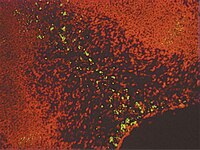
Microglial phagocytosis of living photoreceptors contributes to inherited retinal degeneration.
Zhao, L; Zabel, MK; Wang, X; Ma, W; Shah, P; Fariss, RN; Qian, H; Parkhurst, CN; Gan, WB; Wong, WT
EMBO molecular medicine
7
1179-97
2015
Show Abstract
Retinitis pigmentosa, caused predominantly by mutations in photoreceptor genes, currently lacks comprehensive treatment. We discover that retinal microglia contribute non-cell autonomously to rod photoreceptor degeneration by primary phagocytosis of living rods. Using rd10 mice, we found that the initiation of rod degeneration is accompanied by early infiltration of microglia, upregulation of phagocytic molecules in microglia, and presentation of "eat-me" signals on mutated rods. On live-cell imaging, infiltrating microglia interact dynamically with photoreceptors via motile processes and engage in rapid phagocytic engulfment of non-apoptotic rods. Microglial contribution to rod demise is evidenced by morphological and functional amelioration of photoreceptor degeneration following genetic ablation of retinal microglia. Molecular inhibition of microglial phagocytosis using the vitronectin receptor antagonist cRGD also improved morphological and functional parameters of degeneration. Our findings highlight primary microglial phagocytosis as a contributing mechanism underlying cell death in retinitis pigmentosa and implicate microglia as a potential cellular target for therapy. | | 26139610
 |
Lack of association of C-Met-N375S sequence variant with lung cancer susceptibility and prognosis.
Shieh, JM; Tang, YA; Yang, TH; Chen, CY; Hsu, HS; Tan, YH; Salgia, R; Wang, YC
International journal of medical sciences
10
988-94
2013
Show Abstract
Previously, we identified a sequence variant (N375S) of c-Met gene, however, its association with lung cancer risk and prognosis remain undefined.We investigated the genotype distribution of the c-Met-N375S sequence variant in 206 lung cancer patients and 207 non-cancer controls in the Taiwanese population by DNA sequencing.Lung cancer patients with variant A/G and G/G genotypes showed 1.08-fold increased cancer risk when compared to patients with the wild-type A/A genotype (95% CI, 0.60-1.91). There were no significant differences in postoperative survival between c-Met-N375S and wild-type patients. In the cell model, the c-Met-N375S cells showed a decrease in cell death upon treatment with MET inhibitor SU11274 compared to wild-type cells.Our data suggest that the c-Met-N375S sequence variant may not play a significant role in cancer susceptibility and the prognosis of lung cancer patients. The correlation with chemoresponse of c-Met-N375S is worth further investigation in patients receiving MET therapy. | | 23801885
 |
Metabolism and functions of phosphatidylserine.
Vance, Jean E and Steenbergen, Rineke
Prog. Lipid Res., 44: 207-34 (2005)
2005
Show Abstract
Phosphatidylserine (PS) is a quantitatively minor membrane phospholipid that is synthesized by prokaryotic and eukaryotic cells. In this review we focus on genes and enzymes that are involved in PS biosynthesis in bacteria, yeast, plants and mammalian cells and discuss the available information on the regulation of PS biosynthesis in these organisms. The enzymes that synthesize PS are restricted to endoplasmic reticulum membranes in yeast and mammalian cells, yet PS is widely distributed throughout other organelle membranes. Thus, mechanisms of inter-organelle movement of PS, particularly the transport of PS from its site of synthesis to the site of PS decarboxylation in mitochondria, are considered. PS is normally asymmetrically distributed across the membrane bilayer, thus the mechanisms of transbilayer translocation of PS, particularly across the plasma membrane, are also discussed. The exposure of PS on the outside surface of cells is widely believed to play a key role in the removal of apoptotic cells and in initiation of the blood clotting cascade. PS is also the precursor of phosphatidylethanolamine that is made by PS decarboxylase in bacteria, yeast and mammalian cells. Furthermore, PS is required as a cofactor for several important enzymes, such as protein kinase C and Raf-1 kinase, that are involved in signaling pathways. | | 15979148
 |
The role of phosphatidylserine in recognition and removal of erythrocytes.
Kuypers, F A and de Jong, K
Cell. Mol. Biol. (Noisy-le-grand), 50: 147-58 (2004)
2004
Show Abstract
During the time that erythrocytes (RBC) spend in the circulation, a series of progressive events take place that lead to their removal and determine their apparent aging and limited survival. In addition, a fraction of RBC precursors will be removed during erythropoiesis by apoptotic processes, often described as "ineffective erythropoiesis". Both will determine the survival of erythroid cells and play an important role in red cell pathology, including hemoglobinopathies and red cell membrane disorders. The loss of phospholipid asymmetry, and the exposure of phosphatidylserine (PS) on the surface of plasma membranes may be a general trigger by which cells, including aging RBC and apoptotic cells, are removed. Oxidant stress and inactivation of the system that maintains phospholipid asymmetry play a central role in the events that will lead to PS exposure, death and removal. | | 15095785
 |
Early membrane events in polymorphonuclear cell (PMN) apoptosis: membrane blebbing and vesicle release, CD43 and CD16 down-regulation and phosphatidylserine externalization.
Nusbaum, P, et al.
Biochem. Soc. Trans., 32: 477-9 (2004)
2004
Show Abstract
CD43 down-regulation during the apoptosis of PMN (polymorphonuclear cells) is not caused by proteolysis or internalization. Could it be released with bleb-derived membrane vesicles? Membrane blebbing was followed by microscopy on PMN 'synchronized' by an overnight incubation at 15 degrees C before their spontaneous apoptosis at 37 degrees C. Released vesicles were quantified by flow cytometry. Membrane blebbing, release of bleb-derived membrane vesicles, decrease of CD43/CD16 expression and phosphatidylserine externalization occurred simultaneously. However, caspase and PKC inhibition prevented annexin binding but not blebbing, vesicle release or CD43 expression decrease; myosin light chain kinase inhibition prevented cell blebbing and vesicle release but had no effect on CD43/CD16 down-regulation or annexin V binding. By electron microscopy, CD43 appeared poorly expressed on membrane blebs and concentrated at bleb 'necks'. In conclusion, CD43 down-regulation is not caused by cell blebbing. Cell blebbing, phospholipid 'flip-flop' and CD43/CD16 down-regulation are independent membrane events. | | 15157165
 |
Phosphatidylserine receptor and apoptosis: consequences of a non-ingested meal.
Botto, Marina
Arthritis Res. Ther., 6: 147-50 (2004)
2004
| | 15225357
 |
Hide and seek: the secret identity of the phosphatidylserine receptor.
Williamson, Patrick and Schlegel, Robert A
J. Biol., 3: 14 (2004)
2004
Show Abstract
Phosphatidylserine on the dying cell surface helps identify apoptotic cells to phagocytes, which then engulf them. A candidate phagocyte receptor for phosphatidylserine was identified using phage display, but the phenotypes of knockout mice lacking this presumptive receptor, as well as the location of the protein within cells, cast doubt on the assignment of this protein as the phosphatidylserine receptor. | | 15453906
 |
Exposure of platelet membrane phosphatidylserine regulates blood coagulation.
Lentz, Barry R
Prog. Lipid Res., 42: 423-38 (2003)
2003
Show Abstract
This article addresses the role of platelet membrane phosphatidylserine (PS) in regulating the production of thrombin, the central regulatory molecule of blood coagulation. PS is normally located on the cytoplasmic face of the resting platelet membrane but appears on the plasma-oriented surface of discrete membrane vesicles that derive from activated platelets. Thrombin, the central molecule of coagulation, is produced from prothrombin by a complex ("prothrombinase") between factor Xa and its protein cofactor (factor V(a)) that forms on platelet-derived membranes. This complex enhances the rate of activation of prothrombin to thrombin by roughly 150,000 fold relative to factor X(a) in solution. It is widely accepted that the negatively charged surface of PS-containing platelet-derived membranes is at least partly responsible for this rate enhancement, although there is not universal agreement on mechanism by which this occurs. Our efforts have led to an alternative view, namely that PS molecules bind to discrete regulatory sites on both factors X(a) and V(a) and allosterically alter their proteolytic and cofactor activities. In this view, exposure of PS on the surface of activated platelet vesicles is a key regulatory event in blood coagulation, and PS serves as a second messenger in this regulatory process. This article reviews our knowledge of the prothrombinase reaction and summarizes recent evidence leading to this alternative viewpoint. This viewpoint suggests a key role for PS both in normal hemostasis and in thrombotic disease. | | 12814644
 |
Apoptosis: giving phosphatidylserine recognition an assist--with a twist.
Fadok, Valerie A and Henson, Peter M
Curr. Biol., 13: R655-7 (2003)
2003
| | 12932346
 |
Copper chelation represses the vascular response to injury
Mandinov, L., et al
Proc Natl Acad Sci U S A, 100:6700-5 (2003)
2003
| Immunohistochemistry (Tissue) | 12754378
 |


