Millipore Sigma Vibrant Logo

ABE2984-100UG Anti-Phospho-RXRα/RXRA (Ser56)

NewABE2984-100UG
100 μg  
Retrieving price...
Price could not be retrieved
Minimum Quantity is a multiple of
Maximum Quantity is
Upon Order Completion More Information
You Saved ()
 
Request Pricing
Limited Availability
Stocked 
Discontinued
Limited Quantities Available
Available
    Remaining : Will advise
      Remaining : Will advise
      Will advise
      Contact Customer Service
      Contact Customer Service


      Contact Customer Service

      Overview

      Replacement Information
      Description
      Catalogue NumberABE2984-100UG
      DescriptionAnti-Phospho-RXRα/RXRA (Ser56)
      Alternate Names
      • Retinoic acid receptor RXR-alpha
      • Retinoid X receptor alpha
      • Nuclear receptor subfamily 2 group B member 1
      Background InformationRetinoic acid receptor RXR-alpha (UniProt: P19793; also known as Nuclear receptor subfamily 2 group B member 1, Retinoid X receptor alpha, RXR ) is encoded by the RXRA (also known as NR2B1) gene (Gene ID: 6256) in human. Retinoid X receptor alpha (RXR ), a nuclear receptor of transcription factor, controls various physiological and pathological pathways including cellular growth, proliferation, differentiation, and apoptosis. RXR-alpha is composed of three domains: a modulating N-terminal domain (AF1 domain), a DNA-binding domain, and a C-terminal ligand-binding domain (AF2 domain). RXR-alpha protein is highly expressed in liver and its expression has also been reported in lung fibroblasts, monocytes, kidney, and brain. RXR is phosphorylated on serine and threonine residues mainly at its N-terminal A/B domain by cyclin-dependent kinase 1 (Cdk1) at the onset of mitosis, triggering its translocation to the centrosome, where phosphorylated-RXR (p-RXR ) interacts with polo-like kinase 1 (PLK1) through its N-terminal A/B domain by a unique mechanism. The interaction promotes PLK1 activation, centrosome maturation, and mitotic progression. Studies show that Cdk1 phosphorylation of Ser56 and Ser70 mediates the interaction between p-RXR and PLK1 during mitosis. Importantly, Ser56 and Ser70 and their surrounding amino acids are highly conserved across different species, suggesting their evolutionarily conserved role in regulating RXR activity. An RXR ligand XS060, which specifically inhibits p-RXR /PLK1 interaction but not RXR heterodimerization, promotes mitotic arrest and catastrophe in a tumor-specific manner. Levels of p-RXR are abnormally elevated in cancer cell lines, during carcinogenesis in animals, and in clinical tumor tissues. (Ref.: Xie, G., et al. (2020). Development Cell. 55(6); 707-722; Xie, G., et al. (2021). STAR Protocols. 2(3); 1-15; Zeng, Z., et al. (2015). Cancer Res. 75(10); 2049-2060.)
      References
      Product Information
      FormatPurified
      PresentationPurified rabbit polyclonal antibody in PBS without azide.
      Quality LevelMQ200
      Applications
      ApplicationAnti-Phospho-RXR /RXRA (Ser56), Cat. No. ABE2984, is a rabbit polyclonal antibody that detects RXR and is tested in Western Blotting, Immunohistochemistry, Flow Cytometry, Dot Blot, Immunocytochemistry, Immunoprecipitation, and Immunofluorescence.
      Key Applications
      • Western Blotting
      • Flow Cytometry
      • Immunocytochemistry
      • Immunohistochemistry
      • Immunoprecipitation
      • Dot Blot
      • Immunofluorescence
      Application NotesTested Applications

      Immunofluorescence Analysis: A representative lot detected RXR in Immunofluorescence application (Xie, G., et al. (2021). STAR Protocols. 2, :1-15).

      Western Blotting Analysis: A representative lot detected RXR in Western Blotting application (Xie, G., et al. (2020). Development Cell. 55, : 707-722).

      Immunoprecipitation Analysis: A representative lot immunoprecipitated RXR in Immunoprecipitation application (Xie, G., et al. (2020). Development Cell. 55, : 707-722).

      Immunocytochemistry Analysis: A representative lot detected RXR in Immunocytochemistry application (Xie, G., et al. (2020). Development Cell. 55, : 707-722).

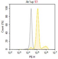
      Flow Cytometry Analysis: A representative lot detected RXR in Flow Cytometry application (Xie, G., et al. (2020). Development Cell. 55, : 707-722).

      Dot Blot: A representative lot detected RXR in Dot Blot application (Xie, G., et al. (2020). Development Cell. 55, : 707-722).

      Flow Cytometry Analysis: 1.0 µg of this antibody detected RXR in HeLa cells overnight serum-starved and treated with Nocodazole 100 ng/ml for 8 hours .

      Immunohistochemistry Applications: A representative lot detected RXR in Immunohistochemistry application (Xie, G., et al. (2020). Development Cell. 55, : 707-722).

      Note: Actual optimal working dilutions must be determined by end user as specimens, and experimental conditions may vary with the end user.
      Biological Information
      ImmunogenKLH-conjugated linear peptide corresponding to 12 amino acids from the surrounding phosphoserine 56 from the N-terminal region of human Retinoic acid receptor (RXR ).
      EpitopeN-terminal
      Concentration0.5 mg/mL. Please refer to guidance on suggested starting dilutions and/or titers per application and sample type.
      HostRabbit
      SpecificityThis rabbit polyclonal antibody detects RXR phosphorylated on serine 56.
      IsotypeIgG
      Species Reactivity
      • Human
      Species Reactivity NoteHuman. Predicted to react with Bovine, Mouse, Rat based on 100% sequence homology.
      Antibody TypePolyclonal Antibody
      Entrez Gene Number
      Gene Symbol
      • RXRA
      Modifications
      • Phosphorylation
      Purification MethodAffinity Purified
      UniProt Number
      Molecular WeightTarget molecular weight ~80 kDa observed; 50.81 kDa calculated. Uncharacterized bands may be observed in some lysate(s).
      Physicochemical Information
      Dimensions
      Materials Information
      Toxicological Information
      Safety Information according to GHS
      Safety Information
      Product Usage Statements
      Quality AssuranceEvaluated by Western Blotting in lysate from HeLa cells in mitotic phase.

      Western Blotting Analysis: A 1:1,000 dilution of this antibody detected RXR phosphorylated on serine 56 in lysate from HeLa cells in mitotic phase.
      Usage Statement
      • Unless otherwise stated in our catalog or other company documentation accompanying the product(s), our products are intended for research use only and are not to be used for any other purpose, which includes but is not limited to, unauthorized commercial uses, in vitro diagnostic uses, ex vivo or in vivo therapeutic uses or any type of consumption or application to humans or animals.
      Storage and Shipping Information
      Storage ConditionsRecommended storage: +2°C to +8°C.
      Packaging Information
      Material Size100 μg
      Transport Information
      Supplemental Information
      Specifications
      Global Trade Item Number
      Catalog Number GTIN
      ABE2984-100UG 04065272074230